

Description
SUMO (Small Ubiquitin-like Modifier) proteins are a family of small proteins that are covalently attached to and detached from other proteins in cells to modify their function. Unlike ubiquitination, which targets proteins for degradation, SUMO modification plays a critical role in a number of cellular functions including nucleocytoplasmic transport, gene expression, cell cycle and formation of subnuclear structures such as promyelocytic leukemia (PML) bodies. There are three confirmed SUMO isoforms in human; SUMO1, SUMO2 and SUMO3. SUMO2 and 3 show a high degree of similarity to each other and are distinct from SUMO1. Individual SUMO family members are all targeted to different proteins with diverse biological functions. SUMO2/3 forms poly-(SUMO) chains, is conjugated to topoisomerase II and APP, and regulates chromosomal segregation and cellular responses to environmental stress.
Molecular mass: SUMO2; proform 10,871 Da with 95 aa (94-95 aa are removed from proform). SUMO3; proform 11,637 Da with 103 aa (93-103 aa are removed from proform).
Applications
- Immunofluorescence staining 1:100-500
- Immunohistochemistry frozen sections 1:100-500
- Western blot 1:1,000
- Immunocytochemistry staining 1:100-500
- ELISA (Assay dependent)
Other applications have not been tested
Specification
Immunogen: Recombinant GST-fused human SUMO3 (full length)
Isotype: Rat IgG 2a kappa
Purification: The antibody was produced from the hybridoma cultured in serum-free medium and purified under mild conditions by propriety chromatography processes.
Form: 1 mg/ml in PBS, 50% glycerol, filter-sterilized. Azide- and carrier protein-free.
Specificity: Specific to human, simian, mouse, hamster and rat SUMO2 and 3. Other species have not been tested.
Storage: Shipped at 4°C or -20°C and store at -20°C
Data Link
Swiss-Prot SUMO2 P61956 (human), SUMO3 P55854 (human)
References
Uchimura Y et al “Involvement of SUMO modification in MBD1- and MCAF1-mediated heterochromatin formation.” J Biol Chem 281: 23180-23190 (2006) PMID: 16757475
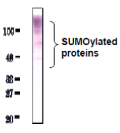
Fig.1. Detection of SUMO-2/3 by Western blot with anti-SUMO2/3 antibody (3H12). High molecular multiple bands were observed in HeLa total cell extract. As secondary antibody, Alkaline phosphatase conjugated anti-rat IgG was used.
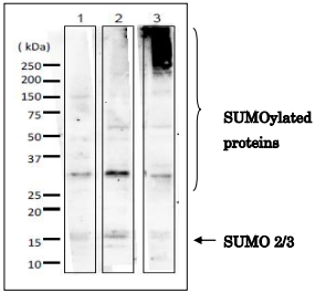
Fig. 2. Detection of SUMO-2/3 in whole cell extracts of mammalian cells by Western blot with anti-SUMO2/3 antibody (3H12). 1. MCF-7 (human breast cancer cell line) 2. NIH3T3 (mouse fibroblast cell line) 3. CHO (Chinese Hamster Ovary cell) 10-20% gradient gel was used for SDS-PAGE. Wet blotting method was employed. Anti-SUMO-2/3 antibody (3H12) was used at 1/1,000 dilution. As a second antibody, goat anti-rat IgG antibody conjugated with HRP was used at 5,000 dilution. Arrow indicates unconjugated SUMO-2/3 proteins. SUMO-2/3 proteins conjugate numerous proteins in vivo, and SUMOylation states vary depending on the kinds of cells and physiological states of them.
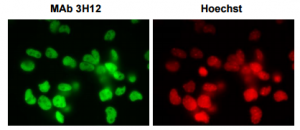
Fig. 3 Immunofluorescence staining of SUMO-2/3 with the anti-SUMO2/3 antibody (3H12) in the mouse primary neural progenitor cells. DNA was stained with Hoechst.
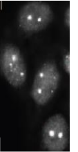
Fig. 4. SUMO-2/3 foci detection in C-33A cells by immunofluorescence staining with anti-SUMO-2/3 antibody (3H12). Cells were fixed with 4% paraformaldehyde and permeabilized with 0.25 TritonX-100. As secondary antibody, Alexa 488 conjugated donkey anti-rat IgG was used. Cells were analyzed using Olympus IX71 microscope and Lumina Vision software (Mitani Co., Tokyo)

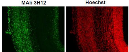
Fig. 5. Immunohistochemistry of Coronal section of E16.mouse cerebral cortex. Coronal section was immunostained with anti-SUMO-2/3 antibody (3H12). DNA was stained with Hoechst.
Reviews
There are no reviews yet.