AS ONE INTERNATIONAL is proud to bring you two brands of distinct hydrogels for 3-D cell culture: Cellendes, a life science company in Germany, developed a biomimetic dextran-based hydrogel while Menicon Life Science in Japan manufactures a peptide-based hydrogel. Each hydrogel offers unique advantages for a variety applications like drug discovery and tissue engineering.
3-D Life Hydrogels from Cellendes
3-D Life hydrogel is a dextran-based hydrogel developed by Cellendes GmbH, a life science company offering a comprehensive technology for controlled design of cell environment in 3D cell culture. The 3-D Life Hydrogel backbone consists of dextran polymer functionalized with maleimides that are cross linked with thiol-bearing linear linkers, PEG-Link or CD-Link. The maleimide-dextran polymer and PEG-Link or CD-Link are mixed together at varying concentrations to adjust the elasticity of the gel. A soft gel may have an elastic shear modulus of 20 Pa while hard gels can reach 6,000 Pa. Hydrogels keep their elasticity at warmer and cooler temperatures and thus can be utilized in a wide range of applications.
3-D Life Hydrogels provide a neutral background matrix that is ideal for incorporating, suspending, and manipulating cells, tissues, biochemicals, and other entities in a “solid” aqueous-based environment or for creating a biologically inert elastic surface or coating. A toolbox of reagents provides flexibility in composing biomimetic hydrogels right at the researcher’s bench. Areas of application include basic research, drug development and biomedical engineering.
3-D Life Hydrogels are biocompatible allowing cells to be embedded during gel formation. Adhesion peptides can be incorporated into the hydrogel to adhere to your cell receptor. Dextranase added to cell culture medium or PBS will dissolve the hydrogel, releasing the cells. Cell integrity and physiology remain functional while in the gel and after gel dissolution.
Gel Characteristics
- Adjustable elasticity– shear modulus from 20 – 6,000 Pa
- High elasticity and low viscosity
- Malleable – can be molded into any shape
- Automation – aqueous solutions of hydrogel components allow microliter precision liquid handling
- Rapid gelation keeps entities suspended in 3-D space
- Scalability – from free-form gels to high throughput applications
- Ambient temperature– gelation occurs at room temperature, no heating or cooling necessary
- Translucent and biocompatible
- Choice of biologically inert or biomimetically modified components
Structure
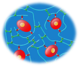
| Maleimide-modified dextran polymer is the biologically inert basis for 3-D Life dextran hydrogels | |
| Crosslinkers PEG or matrix metalloprotease cleavable CD-Link react with maleimide groups on the dextran polymer. Crosslinks polymers to form the gel. | |
| Adhesion peptides (e.g. RGD Peptide) bind via their terminal cysteine to a portion of the maleimide groups of the dextran. Provides a cell adhesion matrix. | |
| Cells bind to the dextran-peptide conjugates with their corresponding adhesion receptors (e.g. integrins) |
Features and Benefits
| Features | Benefits |
| Defined composition of biologically inert synthetic polymers |
|
| User-controlled modifications (e.g. peptides, proteins) |
|
| Robust gel formation and handling |
|
| Wide range of ligand density (up to 5 mmol/L) |
|
| Tunable gel stiffness |
|
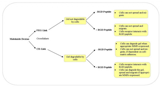
Choosing the right 3-D Life Hydrogel components
Potential Applications
- Suspend biologics or other entities for manipulation
- Suspend chemicals and biochemicals for slow releasing
- Miniaturize your application with microarray hydrogels
- Form an aqueous barrier between different materials or to block an area
- Culture cells in or on top of the hydrogel
- Create an elastic surface
- Create a biocompatible coating to protect tissue sections
- Create a protective coating to reduce sample dehydration
- Automate or create your own cell migration assay
- Drug discovery with 3D cell culture
Application Notes
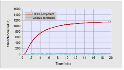
Formation of Hydrogel after mixing of Mal-Dextran and PEG-Link at a final concentration of 3 mMol/L maleimide groups and 3 mMol/L thiol groups.
| Soft gel | Hard gel | |
| Elastic shear modulus | 20 -40 Pa | 4,000 – 6,000 Pa |
| Conc. of thiol groups | 1.5 mMol/L | 6 mMol/L |
| Conc. of maleimide groups | 1.5 mMol/L | 6 mMol/L |
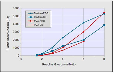
3-D Life Hydrogel components Mal-Dextran and PEG-Link (Dextran-PEG), Mal-Dextran and CD-Link (Dextran-CD), Mal-PVA and PEG-Link (PVA-PEG) and Mal-PVA and CD-Link (PVA-CD), as indicated in the diagram, were mixed together at various final concentrations of reactive groups. The elastic shear modulus of each hydrogel was measured 20 min after the mixing of the polymer components. Each data point represents the average of three independent measurements.
Recent Publications
Ayenehdeh et al. Immunomodulatory and protective effects of adipose tissue-derived mesenchymal stem cells in an allograft islet composite transplantation for experimental autoimmune type 1 diabetes. Immunology Letters. 2017, 188: 21-31.
Martina et al. Modeling human immunity in vitro: improving artificial lymph node physiology by stromal cells. Applied In Vitro Toxicology. 2016, 2(3): 143-150.
Charwat et al. Potential and limitations of microscopy and Raman spectroscopy for live-cell analysis of 3D cell cultures. Journal of Biotechnology. 2015, 205: 70-81.
Order Now:
*Includes maleimide-dextran, crosslinker, buffers, dextranase, thio, water
PanaceaGel from Menicon Life Science
Menicon developed PanaceaGel as a synthetic alternative for collagen scaffolding commonly used in tissue engineering. PanaceaGel is a 13 amino acid peptide that self assembles to form beta sheets. The beta sheets stack on top of each other to form nanofibers. It is the network of interacting nanofibers that form PanaceaGel.
Structure and Mechanism
The main component of PanaceaGel is a thirteen amino acid synthetic peptide, which is made of four naturally occurring amino acids in a specific sequence (US 2012/0058066 A1). The primary structure is:
![]()
The thirteen amino acid peptides undergo spontaneous self-assembly in several steps to form nanofibers in aqueous solvents due to hydrogen bonding, electrostatic interaction and hydrophobic interaction. These nanofibers assemble into a higher-order structure, a nanofiber-scale three-dimensional network, which forms a transparent gel with a moisture content of over 99%.
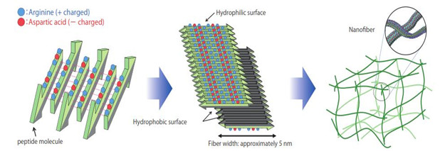
Gel formation by self-assembly
Step 1: Beta sheet structure – 13 amino acid peptides form beta sheets by hydrogen bonding and electrostatic and hydrophobic interactions.
Step 2: Nanofiber formation – Each beta sheet has 2 sides, a hydrophilic surface and a hydrophobic surface. Beta sheets stack on top of each other to form nanofibers in an aqueous solvent (water) by hydrophobic interactions.
Step 3: Nanofiber network structure – Nanofibers interact with each other to form a network containing more than 99% water.
Features and Benefits
| Features | Benefits |
| Self assembly 13 amino acid peptides |
|
| 100% synthetic polymer |
|
| Lot to lot consistency |
|
| Neutral pH |
|
| Stable at room temperature |
|
Recent Publications
Nakamichi et al. Mohawk promotes the maintenance and regeneration of the outer annulus fibrosus of intervertebral discs. Nature Communications, 2016, 7: 12503.
Nagai et al. The mechanical stimulation of cells in 3D culture within a self-assembling peptide hydrogel. Biomaterials, 2012, 33(4): 1044-1051.
Uehara et al. Exogenous fatty acid binding protein 4 promotes human prostate cancer cell progression. Int. J. Cancer, 2014, 135: 2558–2568.
Order Now:
| Cat.# | Component | Size |
| SPG-178-004 | PanaceaGel, 0.4% | 1 ml |
| SPG-178-104 | PanaceaGel, 0.4%, isotonic | 1 ml |
| SPG-178-008 | PanaceaGel, 0.8% | 1 ml |
| SPG-178-108 | PanaceaGel, 0.8%, isotonic | 1 ml |