

Description
Calcium-binding protein that interacts with newly synthesized glycoproteins in the endoplasmic reticulum. It may act in assisting protein assembly and/or in the retention within the ER of unassembled protein subunits. It seems to play a major role in the quality control apparatus of the ER by the retention of incorrectly folded proteins. Associated with partial T-cell antigen receptor complexes that escape the ER of immature thymocytes, it may function as a signaling complex regulating thymocyte maturation. Additionally it may play a role in receptor-mediated endocytosis at the synapse.
Key words: Canx, Endoplasmic Reticulum, Chaperon, Synaptic Vesicle Endocytosis, Transmembrane, Calcium Ion Binding
Molecular mass: 67,278 Da with 591 amino acids. It undergoes palmitoylation and phosphorylation.
Applications:
Western blotting (1/1,000 dilution))
Immunoprecipitation (1/100 dilution).
Immunohistochemistry (1/100 – 1/1,000 dilution)
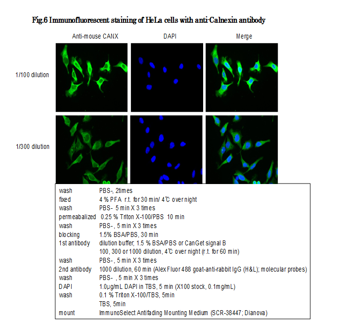
Immuno-fluorescent staining (1/100~1/300 dilution)
Specifications:
Immunogen: C-terminal peptide of mouse Calnexin protein, EDEILNRSPRNRKPRRE, conjugated with KLH
Reactivity: Mouse, rat, human
Form: Whole rabbit antiserum added with 0.1% sodium azide.
Storage: Shipped at 4℃ or at -20℃. Upon arrival, spin-down and store at -20℃ for longer period.
Database Links:
uniprot/P35564 mouse calnexin.
Gene ID 12330 mouse calnexin.
Reference: This antibody was described in Ref.1 and used in the following publications.
Ikawa M. et al. (2001) Calmegin Is Required for Fertilin α/β Heterodimerization and Sperm Fertility. Dev Biol. 240: 254-61. IP. Open access.
Ikawa M. et al. (2011) Calsperin is a testis-specific chaperone required for sperm fertility. J Biol Chem. 286: 5639-46. WB, IP. Open access.
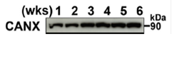
Fig.1 Western blotting analysis of mouse testis extracts of different ages with anti-Calnexin (CANX) antibody. 20 μg of Triton X-100 extracts from mouse testis was reacted with anti-Calnexin antiserum at 1/1,000 dilution. Calnexin expression started at birth. (wks) stands for weeks.
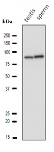
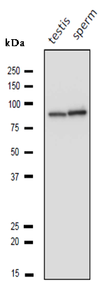
Fig.2 Western blotting analysis of lysatess of mouse testis and sperm with anti-Calnexin (CANX) antibody. Proteins of the lysates (10 μg) of mouse testis and sperm were separated on SDS-PAGE and blotted to PVDF membrane and reacted with anti-Calnexin antiserum at 1/1,000 dilution. Calnexin bands migrated to the position corresponding to molecular mass of 90 kDa.
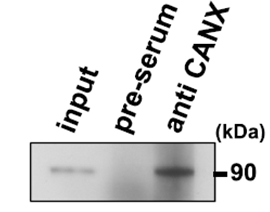
Fig.3.Immunoprecipitation of Calnexin from mouse testis. One mg of testis lysate (supernatant in lysis buffer containing 10 m m Tris-HCl pH8.0, 50 mM NaCl, 1% protease inhibitor mix) was incubated with 2 μl of anti-Calnexin (CANX) antiserum and 50 μl. of protein-A conjugated magnetic beads (Miltenyi Biotec) and immunoprecipitated according to the protocol of supplier. The immunoprecipitated sample was analyzed by western blotting using the same antibody at 1/1,000 dilution.
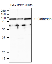
Fig.4 Western blotting analysis of lysatess of human and mouse cell lines with anti-Calnexin (CANX) antibody. Proteins of the lysates (20 μg) of HeLa, MCF7 and NIH3T3 cells were separated on SDS-PAGE and blotted to PVDF membrane and reacted with anti-Calnexin antiserum at 1/1,000 dilution. As second antibody, goat polyclonal antibody to rabbit IgG conjugated with HRP (ab97051) was used.
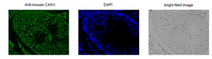
Fig.5. Immunohistochemistry of mouse testis using anti-CANX antibody.
Formalin-fixed and paraffin-embedded mouse testis Deparaffinization by LemosolRA (#122-03991,Wako, Osaka)
Rehydration 100% Et-OH, 95%, 90%, 70%, DW
Antigen retrieval Histo/Zyme (Cat.# k046; Diagnostic BioSystems)
Washing PBST (0.25% triton X-100/PBS-)
Blocking 10 % FBS / PBST 30 min
1st antibody 100 dilution in PBS- 4℃ O/N
Washing PBS-
2nd antibody 1000 dilution, 60 min (AF-488 goat anti-rabbit IgG (H&L),
#1166843 ; molecular probes)
washing PBS- 5 min X 3
DAPI 1.0μg/mL DAPI in TBS 10 min
(×100 stock, 0.1mg/mL)
Washing PBS-
Mount ImmunoSelect Antifading Mounting Medium (SCR-38447; Dianova)
Reviews
There are no reviews yet.