
Description
This is a probable transcription activator that specifically binds the purine-rich single strand of the PUR element located upstream of the MYC gene. Pura may play a role in the initiation of DNA replication and in recombination. Human and mouse Pura has molecular mass of 35 kDa.
Applications
- Western blot 1/1,000~1/3,000 dilution
- Immunoprecipitation 1/1,000 dilution
- Immunofluorescence staining 1/100~1/500
Specifications
Immunogen: Recombinant GST-Pura (human, full-length) expressed in E. coli
Reactivity: Human and mouse. Not tested with other species.
Purification: Affinity-purified from rabbit antiserum with GST-Pura agarose column and anti-GST antibodies were adsorbed with GST agarose column.
Form: 1mg/ml in PBS, 50% glycerol, filter-sterilized. Azide and carrier free.
Shipped at 4°C and stored at -20°C
Data Link
SwissProt: Q00577 Human, Unigene: 443121 Human, Entrez Gene: 5813 Human
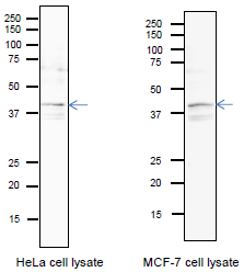
Fig.1 Identification of endogenous Pura protein in whole cell extracts of HeLa cells and MCF-7 cells. Arrow indicates the position of PURA bands in 12.5% SDS-PAGE. Blotting was done in a wet system at 15 volts overnight. The anti-Pura antibody was used at 1/1,000 dilution. CanGetSignal (Toyobo, Osaka) was used as a signal enhancer.
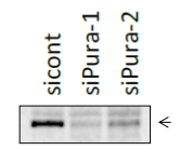
Fig. 2 Identification of Pura protein band by western blot, using siRNA. HCT116 cells were transfected with siPura and the cell lysates were prepared after 48 h. The Pura band was indicated by an arrow at 38 kDa position.
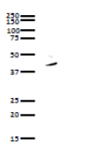
Fig. 3 Immunoprecipitation of Pura protein from whole cell lysate of HeLa cells with anti-PURA antibody. Whole cell lysate of HeLa cells was reacted with anti-Pura antibody and precipitated with protein G conjugated magnetic beads, and analyzed by WB by using anti-Pura antibody. As the secondary antibody, anti-rabbit IgG antibody conjugated with HRP was used.

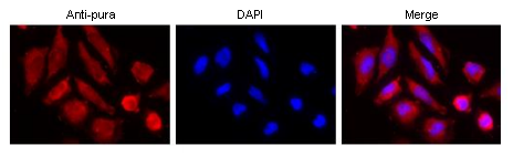
Fig. 4 Immunofluorescence staining of Pura protein in HeLa cells with Anti-Pura antibody. The anti-Pura antibody was used at 1/100 dilution and as the second antibody, Alexa 555-conjugated goat anti-rabbit IgG antibody was used at 1/1,000 dilution. DNA was stained with DAPI.
Reviews
There are no reviews yet.