Description
The Alzheimer amyloid precursor protein (APP) is an integral membrane protein expressed in many tissues and concentrated in the synapses of neurons. Its primary function is not known, though it has been implicated as a regulator of synapse formation and neural plasticity. APP is best known and most commonly studied as the precursor molecule whose proteolysis generates amyloid beta (Aβ), a 39 -42 amino acid peptide whose amyloid fibrillar form is the primary component of amyloid plaques found in the brains of Alzheimer’s disease patients. Isoform APP695 lacking the protease inhibitor domain is the predominant form in neuronal tissues. An antibody (named AN2) against the N-terminus of human APP (aa 18-38) was raised in rabbit.
Applications
Western blot (dilution: 1/3,000-1/1,000)
Immunocytochemistry (dilution: 1/1,000-1/500)
Other applications have not been tested.
Specification
Immunogen: Synthetic peptide corresponding to the N-terminus (aa 18-38) of human APP
Specificity: Specific to human, mouse and rat
Form: Antiserum with 0.05% sodium azide
Storage: Shipped at 4°C and stored at -20°C
Data Link:
UniProtKB/Swiss-Prot P05067 (A4_HUMAN)
References: This antibody was used in ref.3 and 4.
Kang HG et al. (1987) “The precursor of Alzheimer’s disease amyloid A4 protein resembles a cell-surface receptor.” Nature 325: 33-736 PMID: 2881207
Selkoe DJ (1994) “Normal and abnormal biology of the beta-amyloid precursor protein.” Annu. Rev. Neurosci. 17: 489-517 PMID: 8210185
Nishimura I et al. (2002) “Cell death induced by a caspase-cleaved transmembrane fragment of the Alzheimer amyloid precursor protein.” Cell Death Differ. 9: 199-208 PMID: 11840170
Nishimura I et al. (2003) “Upregulation and antiapoptotic role of endogenous Alzheimer amyloid precursor protein in dorsal root ganglion neurons.” Exp. Cell Res. 286: 241-251 PMID: 12749853

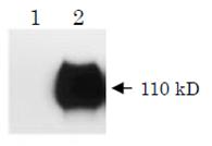
Fig.1 Western blot analysis of APP. Human NT2 neurons infected with adenovirus expressing b-galactosidase (lane 1) or wild-type APP (lane 2) were analyzed by Western blotting using this antibody. Wild-type APP was abundantly expressed in NT2 cells (ref. 3).

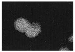
Fig.2 Immunocytochemistry for APP. Mouse dorsal root ganglion cells were treated with this antibody to examine neuronal APP expression (ref. 4).


Reviews
There are no reviews yet.