
Description
ATF6 (activating transcription factor 6) is an endoplasmic reticulum (ER) membrane-bound transcription factor activated in response to ER stress. When unfolded proteins accumulate in the ER, ATF6 is cleaved by regulated intramembrane proteolysis. The resulting amino-terminal fragment translocates to the nucleus and activates transcription by binding to ER stress-response elements present in the promoter regions of ER stress-inducible genes including those encoding ER chaperones and components of ER-associated degradation. ATF6 consists of two closely related factors, ATF6α and ATF6β, in mammals. ATF6α but not ATF6β plays a pivotal role in transcriptional control. The antibody was produced from hybridoma cultured in serum-free medium and purified under mild conditions by propriety chromatography processes.
Applications
Western blotting
Immunoprecipitation (IP) (Less efficient than clone 1-7)
This antibody does not work for immunofluorescence analyses.
Specifications
Immunogen: Recombinant ATF6α (His-tagged amino-terminal fragment of ATF6α fused to GST)
Epitope: Not determined
Isotype: Mouse IgG1 κ
Form: Purified monoclonal antibody (IgG) 1 mg/ml in PBS, 50% glycerol, filter-sterilized
Specificity: Reactive to human and mouse ATF6α. However clone 1-7 antibody (catalog # 73-500) is recommended for human cells.
Storage: -20°C (long period, -70°C)
Data Link:
UniProtKB/Swiss-Prot P18850 (ATF6A_HUMAN)
References
Hai T et al “Transcription factor ATF cDNA clones: an extensive family of leucine zipper proteins able to selectively form DNA-binding heterodimers” Genes Dev 3: 2083-2090 (1989) PMID 2516827
Haze K et al “Mammalian transcription factor ATF6 is synthesized as a transmembrane protein and activated by proteolysis in response to endoplasmic reticulum stress” Mol Biol Cell 10: 3787-3799 (1999) PMID: 10564271
Yamamoto K et al “Transcriptional induction of mammalian ER quality control proteins is mediated by single or combined action of ATF6α and XBP1” Dev Cell 13: 365-376 (2007) PMID: 17765680
Mori K “Divest yourself of a preconceived idea: transcription factor ATF6 is not a soluble protein!”Mol Biol Cell 21: 11435-8 (2010) PMID:20219975
Protocol for ATF6α analysis using anti-human ATF6α monoclonal antibody (37-1)
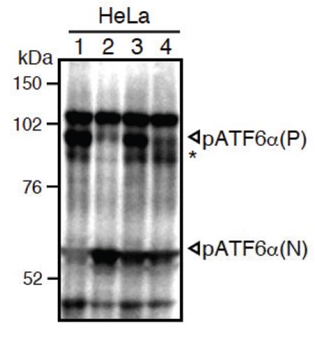
Both endogenous precursor ATF6α, pATF6α(P), and its cleaved product, pATF6α(N), can be detected in human cells such as HeLa cells by western blot analysis using anti-human ATF6α monoclonal antibody clone 37-1 (Fig. 1), according to the procedures described below.
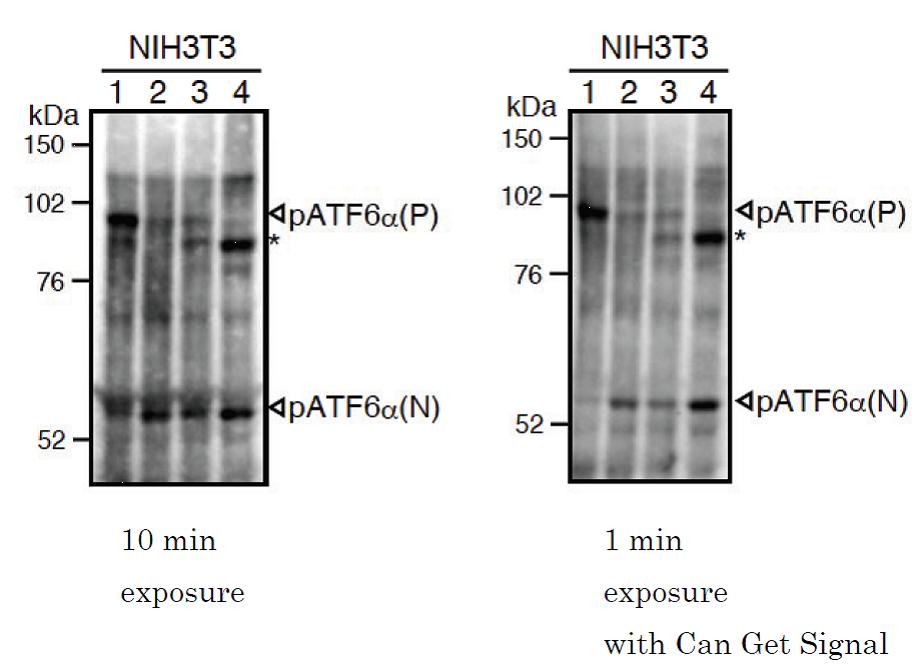
As clone 37-1 cross reacts with mouse ATF6α, both endogenous precursor ATF6α, pATF6α(P), and its cleaved product, pATF6α(N), can be detected in mouse cells such as NIH3T3 cells by western blot analysis (Fig. 2), according to the procedures described below.
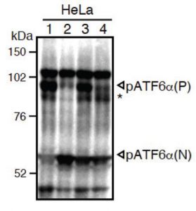
Fig.1 Western blot analysis of human cell extracts using this antibody: Conversion of pATF6α(P) to pATF6α(N) in DTT- or tunicamycin-treated cells.
1) untreated
2) DTT: 1mM dithiothreitol (reducing reagent) for 1 hour.
3) Tm: 2 μg/ml tunicamycin (inhibitor of N-glycosylation) for 3 hour.
4) Tm: 2 μg/ml tunicamycin (inhibitor of N-glycosylation) for 7 hour.
* unglycosylated form of pATF6α (P).
ATF6α is constitutively expressed as pATF6α(P) (~90-kDa protein), and converted to pATF6α(N) (>50-kDa protein) in ER-stressed cells.
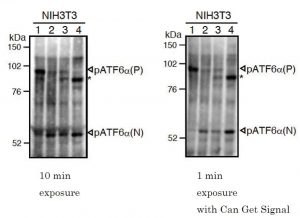
Fig.2 Western blot analysis of mouse cell extracts using this antibody: Conversion of pATF6α(P) to pATF6α(N) in DTT- or tunicamycin-treated cells.
1) untreated.
2) DTT: 1mM dithiothreitol for 1 hour.
3) Tm: 2 μg/ml tunicamycin for 3 hour.
4) Tm: 2 μg/ml tunicamycin for 7 hour.
* unglycosylated form of pATF6α (P).
ATF6α is constitutively expressed as pATF6α(P) (~90-kDa protein), and converted to pATF6α(N) (>50-kDa protein) in ER-stressed cells.
Western blotting
SDS-sample buffer: 50 mM Tris-HCl, pH6.8, containing 2% SDS (100 mM DTT), 10% glycerol and Bromophenol blue
PBST: PBS containing 0.1% Tween 20
Blocking buffer: PBS containing 0.1% Tween 20 and 5% skim milk
Sample Preparation (for HeLa or NIH3T3 cells cultured in a 6 cm dish)
Wash cells with ice-cold PBS.
Scrape cells in 500 μl of ice-cold PBS (+ protease inhibitor cocktail and 10 μM MG132) 2 times and collect cells by centrifugation at 5,000 rpm for 2 min.
Lyse cells directly in 100 µl of SDS-sample buffer without reducing reagent (+ protease inhibitor cocktail and 10 μM MG132).
Vortex vigorously.
Boil the lysate for 5 min, then vortex to mix.
If the lysate is still viscous, boil again and vortex vigorously.
Centrifuge at 14,000 rpm for 2 min.
Determine protein concentration using Bicinchoninic acid protein assay.
SDS-PAGE and incubation with antibody
Add one-tenth volume of 1 M DTT and boil for 5 min.
Use 50 μg of the lysate for an 8% SDS-PAGE.
Transfer to nitrocellulose membrane (such as Hybond-ECL, GE Healthcare).
Incubate the membrane in Blocking buffer overnight at 4°C. Overnight incubation is essential.
Incubate the membrane with primary antibody diluted in Blocking buffer (1:500-1:1000) for 1 hour at room temperature or overnight at 4°C. Wash the membrane 3 times each for 5 min with PBST.
Incubate the membrane with HRP-conjugated secondary antibody for 1 hour at room temperature. We recommend “ECL anti-mouse IgG, Horseradish Peroxidase linked F(ab’)2 fragment” (GE Healthcare NA9310V-1ML).
Wash the membrane 3 times each for 5 min with PBST.
Detect signals using an appropriate luminescent reagent.
*Clearer results can be obtained by using ‘Can Get Signal (TOYOBO NKB-101T)’ during incubation with primary and secondary antibodies, according to the manufacture’s instructions.
Reviews
There are no reviews yet.