

Description
CALR3 capacity for calcium-binding may be absent or much lower than that of CALR. During spermatogenesis, CALR3 may act as a lectin-independent chaperone for specific client proteins such as ADAM3. Required for sperm fertility.
Key words: Calreticulin-3, CALR3, Calsperin, EIF2 complex, Spermatogenesis, Differentiation, Endoplasmic reticulum
Molecular mass: 44,232 with 380 amino acids.
Applications:
Western blotting (1/1,000 dilution))
Immunoprecipitation (1/100 dilution)
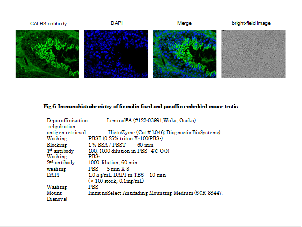
Immunohistochemistry (1/100 dilution)
Other applications have not been tested.
Specifications:
Immunogen: Synthetic peptide corresponding to C-terminal region of mouse CALR3, CMGKFHRHNHLSRFHRQGEL.
Reactivity: Mouse. Not tested with other species.
Form: Rabbit antiserum added with 0.1% sodium azide
Storage: Shipped at 4℃ or -20℃. Upon arrival, spin-down and store at -20℃.
Data Links:
uniprot/Q9D9Q6 mouse
Gene ID 73316 mouse
Reference: This antibody was described and used in the following publication.
Ikawa M. et al (2011) Calsperin is a testis-specific chaperone required for sperm fertility. J Biol Chem.18:5639-46. pubmed/21131354 WB, IP, IHC. Free article.
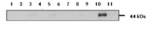
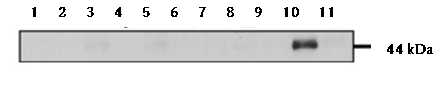
Fig. 1. Testeis specific expression of Calreticulon-3 as examined in various tissues by western blotting with anti-CALR3 antibody. The various tissues were excised and homogenized in lysis buffer containing 1% TritonX100 and then placed on ice for 1 h. These extracts were centrifuged, and the supernatants were collected and analyzed by western blotting with anti-CALR3 antibody at 1/1,000 dilution. 1.Brain. 2. Lung. 3. Heart. 4. Thymus. 5. Liver. 6. Spleen. 7. Kidney. 8. Muscle. 9. Ovary. 10. Testis. 11. Sperm
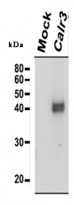
Fig.2. Identification of Calreticulon-3 protein by western blotting with anti-CALR3 antibody. Embryonic fibroblast cells prepared from Calr3 -/- mouse were transfected with a plasmid expressing Calr3. The cell lysate was analyzed by western blotting with anti-CALR3 antibody at 1/1,000 dilution. 1.Mock-infected cell lysate as a negative control; 2.Cell lysate transfected with a plasmid expressing
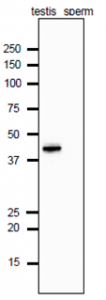
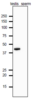
Fig.3. Analysis of Calrecticulon-3 protein in the extracts of mouse testis and sperm by western blotting with anti-Calrecticulon-3 antibody. Proteins in the extracts (10 μg protein) were separated on SDS-PAGE (10-20% gradient gel), electro-blotted to PVDF membrane and reacted with anti-Calrecticulon-3 antibody at 1/1,000 dilution. As the second antibody, anti-rat IgG antibody conjugated with HRP (ab97051) was used at 1/10,000 dilution. The numbers on the left are positions of protein size markers in kDa.
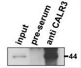
Fig.4. Immunoprecipitation of Calreticulon-3 protein with anti-CALR3 antibody. Lysates of wild-type mouse testis were immunoprecipitated with anti-CALR3 antibody and the precipitates were analyzed by western blotting with the same antibody. 1.Input testis lysate;2.Precipitated with preimmune serum; 3.Precipitated with anti-CALR3 antibody
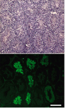
Fig.5. Immunofluorescence staining of a testicular section with anti-CALR3 antibody. Sequential sections were stained with hematoxylin and eosin (upper panel). CALR3 was detected in elongating spermatids (lower panel). Testis was collected from adult mouse and fixed in 4% paraformaldehyde/PBS overnight at 4 °C, cryopreserved in graded 10–30% sucrose, and embedded. Frozen sections (8 μm) were mounted on aminopropyltriethoxysilane-coated glass slides. Primary antibody was used at 1/100 and as secondary antibody, AF-488 conjugated goat anti-rabbit IgG was used. Scale bar is 200 μm.
Reviews
There are no reviews yet.