

Description
CD40 is a 45-50-kDa glycoprotein belonging to the tumor necrosis factor (TNF) receptor superfamily. CD40 is specifically expressed on the surface of B cells and specialized antigen-presenting cells such as dendritic cells and macrophages. CD40 interacts with the CD40 ligand (CD154) which is found primarily on T cells, playing a role in both humoral and cell-mediated immune responses. Activation of CD40 on B cells by CD40 ligand causes B cell proliferation, differentiation, immunoglobulin isotype switching, germinal center formation, and stimulation of the humoral memory response.
This antibody reacts with a 45-48 kDa type I integral membrane glycoprotein present on peripheral blood and tonsillar B cells, but not expressed on terminally differentiated B cells.
The antibody against human CD40 was produced from hybridoma (5C3) cultured in serum-free medium and purified under mild conditions by proprietary chromatography processes.
Applications:
Flow cytometry
Immunohistochemistry (acetone-fixed frozen section)
Immunofluorscence staining 1:10-100
Specifications
Isotype: Mouse IgG1κ
Immunogen: Recombinant extracellular domain of CD40
Conjugate: [FITC] / [IgG] = 5.5
Form: 1.0 mg/ml in PBS, 50% glycerol, filter-sterilized
Specificity: Human
Storage: Shipped at 4°C and stored at -20°C
Data Link:
Swiss-Prot P25942
References:
Inui S et al (1990) Eur J Immunol 20: 1747-1753 PMID: 1698631
Yasui T et al (2002) Int Immunol 14: 319-329 PMID: 11867568
Ishida I et al (2003) Int Immunol. 15: 1027-1034 PMID: 12882840
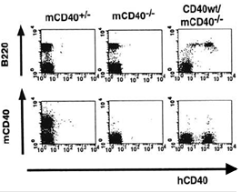
Fig. 1 Flow-cytometry analysis of human CD40 expression in transgenic mouse. Splenocytes from m (mouse) CD40+/- , mCD40-/- and hCD40 wild type/mCD40-/- mice were stained with monoclonal antibodies against mCD40, B220 and hCD40 (5C3) and analyzed by flow cytometry. hCD40 molecules were expressed specifically on B220+ B cells.
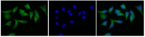
Fig. 2 Immunofluorescence staining of CD40 in MCF7 cells with FITC-conjugated anti-CD40 antibody. Cells were fixed with 4% paraformaldehyde, permeabilized with 0.25% Triton X-100 and stained with FITC-conjugated anti-CD40 antibody (5CS) at 1/20 dilution (left). Nuclear DNA was stained with DAPI (middle). Merged image is shown on right.
Reviews
There are no reviews yet.