
Description
CDT2 (human, 730 aa, 79.5 kDa) is substrate-specific adapter of a DCX (DDB1-CUL4-X-box) E3 ubiquitin-protein ligase complex required for cell cycle control, DNA damage response and translesion DNA synthesis. The DCX(DTL) complex, also named CRL4(CDT2) complex, mediates the polyubiquitination and subsequent degradation of CDT1 and CDKN1A/p21(CIP1). CDT1 degradation in response to DNA damage is necessary to ensure proper cell cycle regulation of DNA replication. CDKN1A/ p21(CIP1) degradation during S phase or following UV irradiation is essential to control replication licensing. Most substrates require their interaction with PCNA for their polyubiquitination: substrates interact with PCNA via their PIP-box, and those containing the ‘K+4’ motif in the PIP box, recruit the DCX(DTL) complex, leading to their degradation. In undamaged proliferating cells, the DCX(DTL) complex also promotes the ‘Lys-164’ monoubiquitination of PCNA, thereby being involved in PCNA-dependent translesion DNA synthesis. CDT2 is activated by checkpoint kinase ATR following DNA damage.
Specification
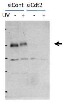
Validation: Specificity was validated by western blotting with siRNA (Fig.2)
Storage: Ship at 4℃, and aliquot and store at -20℃
Reactivity: Human and mouse, rat, hamster
Immunogen: E. coli expressed and purified 6×His-tagged C-terminal 150 amino acids of human Cdt2
Form: Rabbit polyclonal antiserum added with 0.05 % sodium azide
Key words: Ubiquitination, Protein degradation, Cdt1, CDKN1A/p21(CIP1), Cell cycle, DNA damage response, ATR, Phosphorylation, Translesion DNA synsthesis
Applications:
Western blotting (1/200-1/2,000 dilution).
Immunoprecipitation (assay dependent)
Immunofluorescence staining (1/100~1/1,000 dilution)
Flow Cytometry (assay dependent).
Database links:
SwissProt: Q9NZJ0 Human;
Entrez Gene: 51514 Human
References:
This product was described in Ref.1 and used in the following publications.
Nishitani H. et al. (2008) CDK Inhibitor p21 Is Degraded by a Proliferating Cell Nuclear Antigen-coupled Cul4-DDB1Cdt2Pathway during S Phase and after UV Irradiation. J.Biol.Chem. 283: 29045-29052. WB Link
Ishii T. et al. (2010) Proliferating cell nuclear antigen-dependent rapid recruitment of Cdt1 and CRL4Cdt2 at DNA-damaged sites after UV irradiation in HeLa cells. J Biol Chem. 285:41993-42000. WB, IF, FACS Link
Roukos V. et al. (2011) Dynamic recruitment of licensing factor Cdt1 to sites of DNA damage. J. Cell Science 124: 422-434. IF Link
Sakaguchi H. et al. (2012) Checkpoint Kinase ATR Phosphorylates Cdt2, a Substrate Receptor of CRL4 Ubiquitin Ligase, and Promotes the Degradation of Cdt1 following UV Irradiation. PLoS ONE 7(9): e46480. WB Link
Shiomi Y. et al. (2012) Two Different Replication Factor C Proteins, Ctf18 and RFC1, Separately Control PCNA-CRL4Cdt2-Mediated Cdt1 Proteolysis during S Phase and following UV Irradiation. Mol. Cell. Biol. 32: 2279-2288. WB, IF Link
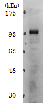
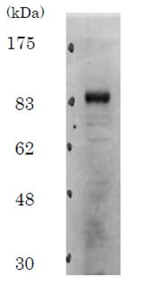
Fig.1. Identification of Cdt2 protein in whole cell extract of HeLa cells by western blotting. With cell extract of 20 μg protein, endogenous level of Cdt2 protein was detected with the antibody at 1/2,000 dilution. Secondary antibody was HRP-conjugated goat anti-rabbit IgG antibody at 1/20,000.
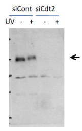
Fig.2. Inhibition of Cdt2 protein synthesis by Cdt2 siRNA introduced into HeLa cells. siCont is control siRNA unrelated to Cdt2. siCdt2 is Cdt2 specific siRNA. Cdt2 is phosphorylated after UV irradiation as shown by the band shift-up in irradiated sample (UV +).
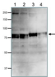
Fig.3. Detection of endogenous Cdt2 protein in whole cell extracts of human and rodent cells by western blotting. Samples, 40 μg, applied. The antibody used at 1/200 dilution. Doublet bands reflect post-translational phospholylation. 1. HeLa cells (human); 2. MCF7 cells (human); 3. NIH3T3 cells (mouse); 4. CHO cells (hamster)
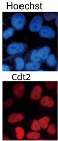
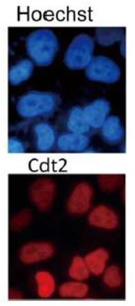
Fig. 4. Immunofluorescence staining of Cdt2 protein in growing Hela cells with the antibody. Asynchroneously growing HeLa cells were stained with Hoechst 33258 for DNA and with the antibody for Cdt2 protein. The cells were fixed in 4% paraformaldehyde for 10 min, permeabilized in 0.25% (v/v) Triton X-100 in phosphate-buffered saline (PBS), and reacted with the primary antibody. AF-592 conjugated goat anti-rabbit IgG was used as secondary antibody. Cdt2 protein is localized in nuclei at all cell stages.
Reviews
There are no reviews yet.