
Description
Cholera toxin, a main enterotoxin, interacts with G proteins and increases cyclic AMP in the intestinal lining to open ion channels. As ions flow into the intestinal lumen (lining), body fluids (mostly water) flows out of the body due to osmosis leading to massive diarrhea as the fluid is expelled from the body. Cholera toxin is a complex consisting of one molecule of A subunit (27.2 kD) and 5 molecules of B subunits (11.6 kD). After secretion, A subunit is proteolytically processed into A1 (22 kD) and A2 (5 kD) subunits which are held together by a disulfide bond. The toxin adsorbs to GM1 ganglioside on the surface of target cells by the B subunit and the A subunit is dissociated from the B subunit during penetration. The A subunit constitutively activates adenyl cyclase activity of α subunit of Gs (a kind of GTP-binding protein).
Applications
Western blot (dilution: 1/2,000)
Immunoprecipitation
Other applications have not been tested.
Specifications
Immunogen: Cholera toxin and the toxoid purified from culture medium of Vibrio cholerae 569B strain
Form: Antiserum added with 0.05% sodium azide
Storage: Shipped at 4°C or -20°C, and upon arrival, aliquot and long term storage -20°C or below
Data Link
UniProt KB Cholera toxin
References:
Hirst TR and D’Souza JM In The Comprehensive Sourcebook of Bacterial Protein Toxins Alouf J and Popoff M edt. 3rd edn. p.270-290 Academic Press (2006)
Finkelstein RA and LoSpalluto JJ “Pathogenesis of experimental cholera. Preparation and isolation of choleragen and choleragenoid.” J. Exp Med 130: 185-202 (1969) PMID: 4978880

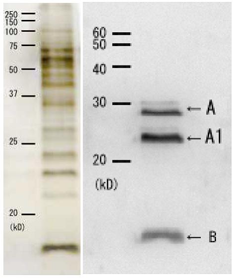
Fig.1 A SDS-PAGE analysis of culture medium of Vibrio cholerae. Culture medium of Vibrio cholerae, 569B strain was subjected to electrophoresis under reducing condition followed by silver-staining.
Fig.1 B Western blotting of culture medium of Cholera toxin.
Culture medium of Vibrio cholerae 569B strain was subjected to electrophoresis under reducing condition followed by Western blotting using this antibody (1/2000 dilution). A, A1 and B indicate the subunits A, A1 and B, respectively. A faint band above A is a precursor of A with signal peptide. A2 subunit (5 kD) is too small to be seen by this analysis.
Reviews
There are no reviews yet.