

Description
Background:
IGSF8 may play a key role in diverse functions ascribed to CD81 and CD9 such as oocytes fertilization or hepatitis C virus function. May regulate proliferation and differentiation of keratinocytes. May be a negative regulator of cell motility: suppresses T-cell mobility coordinately with CD81, associates with CD82 to suppress prostate cancer cell migration, regulates epidermoid cell reaggregation and motility on laminin-5 with CD9 and CD81 as key linkers. May also play a role on integrin-dependent morphology and motility functions. May participate in the regulation of neurite outgrowth and maintenance of the neural network in the adult brain.
Molecular mass: 65,011 Da with 611 amino acids
Specifications:
Reactivity: Mouse. Likely to react with rat and human due to high sequence homology.
Immunogen: Full-length mouse IGSF8 with Flag tag
Form: 0.5 mg/ml IgG fraction of antiserum in PBS, 50% glycerol, 0.05% sodium azide.
Validation: Specificity validated with knock-out mouse (Fig.1)
Storage: Shipped at 4℃and store at -20℃.
Applications:
- Western blotting (1/500~1/1,000 dilution))
- Immunofluorescence and immunochemical staining (1/100 dilution).
- Immunohistochemical staining (1/100)
Data Links: uniprot/Q8R366 mouse IGSF8 Gene ID140559 mouse IGSF8
Reference: This antibody was described and used in the following publication.
- Inoue N. et al Tetraspanin-interacting protein IGSF8 is dispensable for mouse fertility. Fertil Steril. 2012 98(2):465-70.
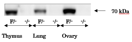
Fig 1. Analysis of IGSF8 protein in various tissues of Igff8-targeted mice by western blotting with anti-IGFS8 antibody. Lysates of tissues (30 μg) were analyzed by western blotting using the antibody at 1/500 dilution. “ F ” and “ – “ stand for floxed and knock-out alleles, respectively.
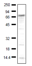
Fig.2 Detection of endogenous level of IGSF8 in crude extract of NIH3T3 cells by using anti-IGSF1 antibody.
Protiens in 40 ug of the cell extract were separated by 12.5% SDSD-PAGE and electro-blotted at 15v, over night (wet system).
Blocking, 1hr, room temp.
1st antibody 1/1000 dilution
2nd, Goat polyclonal secondary antibody to rabbit IgG-H&L (HRP), ab97051
Positions marker proteins are shown in kDa on the left
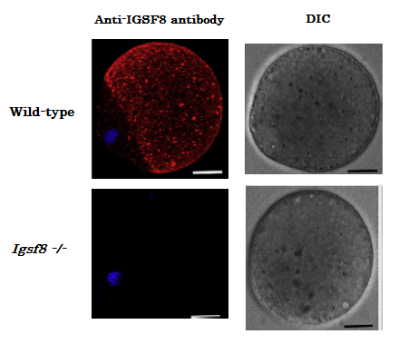
Fig.3. Immunofluorescence staining of IGSF8 protein in eggs of wild-type mouse and Igsf8 knock-out mouse with anti-IGSF8 antibody. Zona-free eggs were fixed in PBS containing 0.5% (v/v) polyvinylpyrrolidone and 4% (v/v) paraformaldehyde. The anti-IGSF8 antibody was used at 1/100 dilution and as the second antibody, Alexa-Fuor 546 labeled anti-rabbit IgG was used (red). Then the DNA was stained with Hoechst 33342 (blue). “DIC” is picture of Differential Interference Contrast microscopy.
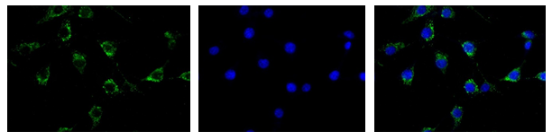
Fig.4. Immunofluorescence staining of IGSF8 protein in NIH3T3 cells with anti-IGSF8 antibody.
NIH3T3 cell were fixed in 4% (v/v) paraformaldehyde. The anti-IGSF8 antibody was used at 1/100 dilution and as the second antibody, Alexa-Fuor 488 labeled anti-rabbit IgG was used (green) at 1/1,000 dilution. DNA was stained with DAPI (blue).
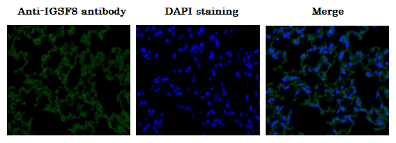
Fig.5 Immunohistochemical staining of IGSF8 protein in mouse lung tissue section using anti-IGSF1 antibody.
4% PFA fixed section of mouse lung tissue
Deparaffinization; LemosolRA (#122-03991, Wako, Osaka)
Rehydration
Antigen retrieval; Histo/Zyme (Cat.# k046; Diagnostic BioSystems)
Washing; PBST (0.25% triton X-100/PBS-)
Blocking: 1 % BSA / PBST 60 min
1st antibody; 1/100 dilution in PBS- 4℃ overnight
Washing; PBS-
2nd antibody; 1/1,000 dilution, 60 min
Washing; PBS-, 5 min 3 times
DAPI; 1.0 μg/mL DAPI in TBS 10 min
Washing; PBS-
Mount: ImmunoSelect Antifading Mounting Medium (SCR-38447; Dianova)
Reviews
There are no reviews yet.