
Description
Background: Matrix protein 1 (M1) plays critical roles in virus replication, from virus entry and uncoating to assembly and budding of the virus particle. M1 binding to ribonucleocapsids (RNPs) in nucleus seems to inhibit viral transcription. Interaction of viral NEP with M1-RNP is thought to promote nuclear export of the complex, which is targeted to the virion assembly site at the apical plasma membrane in polarized epithelial cells. Interactions with NA and HA may bring M1, a non-raft-associated protein, into lipid rafts. Forms a continuous shell on the inner side of the lipid bilayer in virion, where it binds the RNP. During virus entry into cell, the M2 ion channel acidifies the internal virion core, inducing M1 dissociation from the RNP. M1-free RNPs are transported to the nucleus, where viral transcription and replication can take place. M1 of Influenza B virus consists of 248 amino acids with molecular mass of 27,415.
Applications
- Western blotting (1/500~1/1,000 dilution)
- Immunofluorescent and Immunocytochemical staining (1/100~1/200 dilution)
- Immunoprecipitation (1/200 dilution)
- ELISA (assay dependent)
Specification
Immunogen: Human Influenza B Virus strain Nagasaki/1/87, one of the strains of B/Victoria group.
Reactivity: Reacts with M1 proteins of all Influenza B viruses so far tested (100 strains) as examined by ICC staining, including lineage Yamagata strains; Mie/1/1993, Johanesburg/5/1999, Florida/4/2006 and lineage Victoria strains; Lee/1940, Gif/21/1973, Shangdong/7/1997, Malasia/2506/2004, Massachustts/2/2012
No cross reactivity with influenza A viruses.
Isotype: mouse IgG1, kappa
Purity: Produced in serum-free medium and purified by proprietary chromatography procedure under mild conditions.
Form: 1 mg/ml in PBS, 50% glycerol, filter sterilized
Storage: Shipped at 4°C or -20°C. Upon arrival, spin down and store at -20°C.
Data Link: UniProt/A4D3F9 Influenza B virus Matrix 1 protein
References: This antibody was described and used in the following references.
Nakagawa N. et al. Rapid detection and identification of two lineages of influenza B strains with monoclonal antibodies. J Virol Methods. 1999;79:113-2 ICC, IP
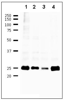
Fig. 1 Detection of M1 protein in the crude extracts of MDCK cells infected with various Influenza B virus strains by using 6A11 monoclonal antibody
- B/Florida/4/2006
- B/Lee/1940
- B/Malasia/2506/2004
- B/Massachusetts/2/2012
First antibody was used at 1/500 dilution and as 2nd antibody, HRP-conjugated goat anti-mouse IgG antibody was used at 1/10,000 dilution. Positions of marker proteins are indicated in kDa on the left.

Fig.2 Immunofluorescence assay of MDCK (canine kidney ) cells infected with Influenza B virus, using anti-Influenza B virus M1 antibody (clone 6A11)
Anti-Influenza B Virus M1 antibody (clone 6A11) efficiently detected the viruses in the infected MDCK cells with B/Florida/4/2006 virus. The cells were fixed with 4% paraformaldehyde in phosphate-buffered saline (PBS) and permeabilized with 0.1% Triton X-100 in PBS. The antibody (6A11) was used at 1/100 ditlution and the bound antibody was visualized by a further reaction with an Alexa Fluor 488-conjugated secondary antibody (green). Image on the left is a negative control, mock-infected MDCK cells.
Reviews
There are no reviews yet.