
Description
Background: Influenza virus nucleoprotein (NP) is a major component of the ribonucleoprotein complex and is abundantly expressed during the course of infection. It is a structural protein, which encapsidates the negative strand viral RNA and is essential for RNA transcription, replication and packaging. From the nucleotide sequence, NP is consists of 560 amino acids with a calculated molecular mass of 61,770.
Post-translational modification: NP may be cleaved from a 56-kDa protein to a 53-kDa protein by a cellular caspase in infected cells. This cleavage may be a marker for the onset of apoptosis in infected cells or have a specific function in virus-host interaction.
Applications
- Immunofluorescent and Immunocytochemical staining (1/100 dilution)
- Immunoprecipitation (1/100 dilution)
- ELISA (assay dependent)
May not suitable for Western blotting
Specification
Immunogen: Human Influenza B Virus strain Nagasaki/1/87, one of the strains of B/Victoria group
Reactivity: Reacted with NP of all Influenza B viruses so far tested (113 clinical strains), including Yamagata lineage strains Mie/1/1993, Johannesburg/5/1999, Florida/4/2006 and Victoria lineage strains Lee/1940, Gif/21/1973, Shangdong/7/1997, Malasia/2506/2004, Massachustts/2/2012
No cross reactivity with influenza A viruses.
Isotype: mouse IgG1, kappa
Product: Produced in serum-free medium and purified by proprietary chromatography procedure under mild conditions.
Form: 1 mg/ml in PBS, 50% glycerol, filter sterilized.
Storage: Shipped at 4°C or -20°C. Upon arrival, spin down and store at -20°C.
Data Link: UniProt/P04665 Influenza B virus nucleoprotein
References: This antibody was described and used in the following references.
Nakagawa N. et al. Rapid detection and identification of two lineages of influenza B strains with monoclonal antibodies. J Virol Methods. 1999;79:113-2 ICC, IP
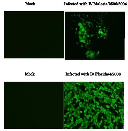
Fig.1 Immunofluorescence assay of MDCK (canine kidney) cells infected with Influenza B virus, using anti-Influenza B virus NP antibody (clone 8C8)
Samples were taken at 24 hours post-infection. Anti-Influenza B Virus NP antibody (clone 8C8) efficiently detected the viruses in the infected MDCK cells. The cells were fixed with 4% paraformaldehyde in phosphate-buffered saline (PBS) and permeabilized with 0.1% Triton X-100 in PBS. The bound antibody was visualized by a further reaction with an Alexa Fluor 488-conjugated secondary antibody. Images on the left are mock-infected MDCK cells as negative control. The cells infected with an Influenza B virus vaccine strain, Malaysia/2506/2004 of Victoria group is shown in the upper panel and Florida/4/2006 of Yamagata group in the lower panel.
Reviews
There are no reviews yet.