
Description
Function: Legionnaires disease (LD) was recognized in 1976 after an outbreak of pneumonia at an American Legion convention in Philadelphia. Soon after, the etiologic agent was identified as a fastidious gram-negative bacillus and named Legionella pneumophila. Although several other species of the genus Legionella were subsequently identified, L pneumophila is the most frequent cause of human legionellosis and a relatively common cause of community-acquired and nosocomial pneumonia in adults. In children, L pneumophila is also an important, although relatively uncommon, cause of pneumonia.
Applications
- Immunofluorescent and Immunochemical staining (1/10,000 ~1/30,000 dilution)
- Immunohistochemistry (1/3,000~1/10,000)
- ELISA (1/10,000~1/30,000 dilution)
- Agglutination (1/2,000~1/5,000)
Specification
Immunogen: Formaldehyde treated whole cells of Legionella pneumophila strain Philadelphia 1 (ATCC #33152). Immunized 7 times at two-week intervals.
Reactivity: Reacts with Legionella pneumophila strains. Since the antiserum has not been adsorbed, it may cross-reacts with related bacteria.
Form: Undiluted antiserum added with 0.09% sodium azide.
Storage: Shipped at 4°C. Upon arrival, aliquot and store at -20°C. Avoid repeated freeze-thaw cycles.
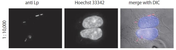
Fig.1 Immunofluorescent staining of Legionella pneumophila in infected HEK293 cells using anti-Legionella pneumophila antibody
HEK293 cells were infected with Legionella pneumophila strain Philadelphia1, fixed with 4% formaldehyde, and reacted with the anti-Legionella pneumophila antibody at 1/10,000 dilution. As a second antibody, goat Rodamine Red X conjugated anti-rabbit IgG antibody was used at 1/10,000 dilution. DNA was stained with Hoechst 33342 (center) and the images were merged with that of differential interference contrast microscope (right).
Reviews
There are no reviews yet.