
Description
MAGE-D1 (melanoma-associated antigen D1), also known as Dlxin-1 or NRAGE, is a member of the MAGE protein family. MAGE-D1 is expressed in almost all normal adult tissues and has been demonstrated to interact with the p75 neutrophin receptor and to facilitate nerve growth factor-dependent apoptosis (ref.1). MAGE-D1 also interacts with its homologous protein, necdin, which is known as a growth suppressor and a promoter of differentiation in neurons. Necdin binds to Msx (msh homeobox) and Dlx (distal-less homeobox) family homeodomain transcription factors via MAGE-D1 and these proteins cooperate to modulate differentiation of neurons and muscle cells by regulating gene expression. Msx proteins function as transcriptional repressor whereas Dlx proteins are transcriptional activators (ref. 2 and 3).
MD1 antibody against mouse MAGE-D1 was raised in rabbit (ref. 2).
Applications
- Western blotting (dilution: 1/3,000-1/1,000)
- Immunohistochemistry (dilution: 1/300)
- Immunocytochemistry (dilution: 1/1,000-1/500)
Not tested for other applications.
Specification
Immunogen: Recombinant MBT-fused mouse MAGE-D1 (aa 1-775 )
Specificity: React with mouse, rat and human MAGE-D1
Form: Antiserum with 0.05% sodium azide
Storage: Shipped at 4°C and stored at -20°C
Data Link: Swiss-Prot Q9QYH6 (mouse), Q9ES73 (rat), Q9Y5V3 (human)
References: This antibody was produced and used in ref. 2 and 3.
- Salehi AH et al (2000) “NRAGE, a novel MAGE protein, interacts with the p75 neutrophin receptor and facilitates nerve growth factor dependent apoptosis” Neuron 27: 279-288 PMID: 10985348
- Kuwajima T et al (2004) “Necdin interacts with the Msx2 homeodomain protein via MAGE-D1 to promote myogenic differentiation of C2C12 cells” J Biol Chem 279: 40484-40493 PMID: 15272023
- Kuwajima T et al (2006) “Necdin promotes GABAergic neuron differentiation in cooperation with Dlx homeodomain proteins” J Neurosci 26: 5383-5392 PMID: 16707790
Related products:
# 74-100 anti-Necdin antibody
# 74-114 anti-MAGE-G1 / Necdin-like 2 antibody
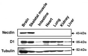
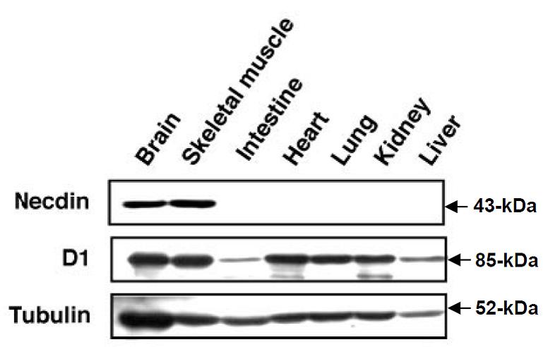
Fig.1 Western blot analysis using this antibody.
Western blot analysis of necdin and MAGE-D1 in mouse embryonal tissues (ref. 2).
Tissue lysates (20 ug) from E18.5 embryos were separated by SDS-PAGE and immunoblotted with antibodies against necdin, MAGE-D1 (D1) and tubulin. Endogenous ~43-kDa necdin protein was detected almost exclusively in the brain and skeletal muscle whereas endogenous ~85-kDa MAGE-D1 protein was expressed in a ubiquitous manner.
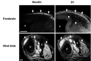
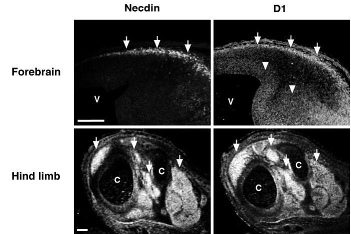
Fig.2 Immunohistochemistry using this antibody (ref. 2).
Fluorescence immunohistochemistry for necdin and MAGE-D1 in mouse embryos.
Adjacent sections of the forebrain at E12.5 embryo (upper panels) and the hind limb at E14.5 (lower panels) were stained with antibodies against necdin and MAGE-D1.
The arrows point to the preplate (upper panels) and the skeletal muscles (lower panels). The arrowheads indicate the ventricular proliferative zone; V, ventricle; C, bone cavity.
Necdin and MAGE-D1 were concentrated in the preplate of the forebrain at E12.5 and skeletal muscle tissues in the hind limb at E14.5. In developing neural tube, MAGE-D1 immunoreactivity was distributed in the ventricular zone as well as the marginal zone.
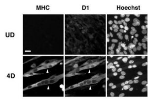
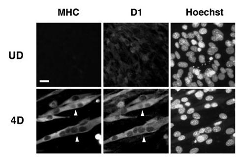
Fig.3 Immunocytochemistry using this antibody (ref. 2).
C2C12 cells were cultured under undifferentiated conditions (UD) or under differentiation conditions for 4 days (4D). Cells were fixed and triply stained for myosin heavy chain (MHC), MAGE-D1 (D1), and nuclear DNA with Hoechst33342 (Hoechst). The arrowheads point to differentiated multinucleated myocytes.
MHC and MAGE-D1 were distributed predominantly in the cytosol of multinucleated differentiated C2C12 cells.
Reviews
There are no reviews yet.