
Description
Description
MCM7 (human; 718 aa, 80 kDa) acts as component of the MCM2-7 complex (MCM complex) which is the putative replicative helicase essential for ‘once per cell cycle’ DNA replication initiation and elongation in eukaryotic cells. The active ATPase sites in the MCM2-7 ring are formed through the interaction surfaces of two neighboring subunits such that a critical structure of a conserved arginine finger motif is provided in trans relative to the ATP-binding site of the Walker A box of the adjacent subunit. The six ATPase active sites, however, are likely to contribute differentially to the complex helicase activity. Required for S-phase checkpoint activation upon UV-induced damage.
This product is purified IgG from the rabbit antiserum.
Applications
- Western blot (1/1,000~1/5,000 dilution).
- Immunoprecipitation (assay dependent)
- Immunofluorescence staining (1/200~1/1,000 dilution).
Other applications have not been tested.
Specification
Immunogen: Purified His6-tagged human MCM7 protein encompassing 562 -719 amino acids.
Reactivity: Reacts with human mouse, rat and hamster. Not tested in other species.
Form: 1 mg/ml in PBS, 50% glycerol, filter-sterilized, sodium azide and carrier-free.
Storage: Ship at 4°C. Upon arrival, centrifuge, aliquot and store at -20°C.
Data Link UniProtKB/Swiss-Prot P33993 MCM7_HUMAN
Reference: This antibody was described and used in the following publications.
- Fujita M et al. (1996) hCDC47, a human member of the MCM family. Dissociation of the nucleus-bound form during S phase. J Biol Chem. 271:4349-54. PMID 8626784 Free Article. WB, IP, IF
- Fujita M. et al. (1997) In vivo interaction of human MCM heterohexameric complexes with chromatin. Possible involvement of ATP. J Biol Chem. 272:10928-35. PMID 9099751 Free Article. WB, IP
- Fujita M. et al. (2002) Nuclear organization of DNA replication initiation proteins in mammalian cells. J Biol Chem. 277:10354-61. PMID 11779870 Free Article. WB, IP, IF.
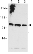
Fig. 1. Identification of MCM7 protein in whole cell extracts of human cells by Western blot using anti-MCM7 antibody.
Lane 1: SiHa cells
Lane 2: C33A cells
Lane3: WI38 cells
All cell lines are cervical cancer derived. Samples are obtained from
approximately 105 cells.
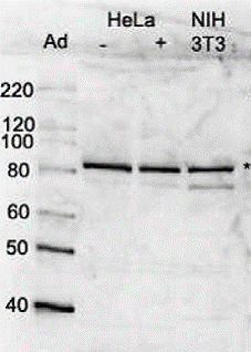
Fig. 2 Identification of MCM7 protein in whole cell extracts of human and mouse cells by Western blot using anti-MCM7 antibody.
Lane 1: Size marker proteins in kDa.
Lane 2: Extract of HeLa cells untreated (-).
Lane 3: Extract of HeLa cells treated with 100 nM adriamycin for 24 hr (+)
Lane 4. Extract of NIH3T3 (mouse) cells.
Anti-MCM7 antibody was used at 1/2,000 dilution.
* Indicates the band of MCM7 protein
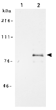
Fig. 3. Immunoprecipitation of MCM7 protein from crude extract of human fibroblast cell line WI38 by using anti-MCM7 antibody.
Lane 1: Immunoprecipitation with pre-immune serum
Lane 2: Immunoprecipitation with anti-MCM7 antiserum.
Cells were labeled with S35 methionine and MCM7 was immunoprecipitated with the anti-MCM7 antibody followed by SDS-PAGE and autoradiography.
Fig. 4. Immunofluorecence staining and confocal microscopic analysis of MCM7 in G1 phase HeLa cell nucleus by using anti-MCM7 antibody after treatment with protein cross-linking reagent, DSP and chromatin extraction. The processed cells were fixed with formaldehyde before staining.
Reviews
There are no reviews yet.