
Description
Mitf (Microphthalmia-associated transcription factor) is a transcription factor that contains both basic helix-loop-helix and leucine zipper structural features. It plays a critical role in the differentiation of various cell types such as neural crest-derived melanocytes, mast cells, osteoclasts and optic cup-derived retinal pigment epithelium. Mutations in Mitf cause auditory-pigmentary syndromes, such as Waardenburg syndrome type 2 and Tietz syndrome. Alternatively spliced transcript variants encoding different isoforms have been identified.
The antibody was produced by immunizing rabbit with recombinant human Mitf protein in the laboratory of Prof. H. Yamamoto.
Applications
- Western blot (1/5,000: Different splicing isoforms detected)
- Immunohistochemistry (dilution: 1/500 ~ 1/1,000)
- Immunocytochemistry
- ChIP (1/200: Users should examine the best conditions which depend on the samples and extract preparation)
Specification
Immunogen: Recombinant full-size human Mitf protein with His tag
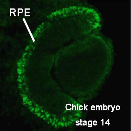
Specificity: Specific to human, mouse, chicken and Xenopus Mitf. Especially it works well with the eye
Form: Antiserum added with 0.05% sodium azide
Storage: -20°C
Data Link: Swiss-Prot : O75030 (human), Q08874 (mouse), O73871 (chicken), A4IID0 (Xenopus), OMIM (human): 156845
References: This antibody was used in the following references.
- Tsukiji N et al “Mitf functions as an in ovo regulator for cell differentiation and proliferation during development of the chick RPE.” Dev Biol 326: 335-346 (2009) PMID: 19100253
- Delmas V et al “beta-Catenin induces immortalization of melanocytes by suppressing p16INK4a expression and co-operates with N-Ras in melanoma development.” Genes Dev21: 2923-2935 (2007) PMID: 18006687
- Osawa M et al “Molecular characterization of melanocyte stem cells in their niche. “Development 132: 5589-5599 (2005) PMID: 16314490
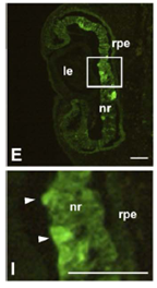
Fig.1 Expression of Mitf protein in wild-type-Mitf-transfected chicken embryo (embryo was harvested 48h after transfection) (ref. 1).
Panel I shows magnifications of the framed area in panel E. rpe, retinal pigment epithelium; nr, neural retina; le, lens. Scale bars = 100 um. Arrowheads indicate the areas where Mitf is strongly expressed.
Reviews
There are no reviews yet.