
Description
Background:
Nucleobindin 2 (NUCB2), also known as NEFA or Nesfatin precursor, is a ubiquitously expressed EF-hand Ca2+ binding protein that is implicated in various physiological processes. Nucleobindin 2 interacts with the postmitotic growth suppressor necdin in neurons. Both necdin and nucleobindin 2 are expressed in differentiated neurons and skeketal muscles and these proteins are likely to be involved in the regulation of survival and death of postmitotic cells by controlling Ca2+ homeostasis.
Specifications:
Reactivity: Reacts with mouse rat and human nucleobindin 2.
Immunogen: Recombinant GST-fused mouse nucleobindin 2 (aa 26-420)
Form: Protein A-affinity purified IgG. 1 mg/ml in PBS, 50% glycerol. Filter-sterilized. No additive.
Storage: Ship at 4℃ and store at -20℃.
Applications:
- Western blotting (1/1,000-1/3,000).
- Immuno-precipitation. (assay dependent)
- Immuno-cytochemistry (1/300-1/1,000)
- Immuno-histochemistry (1/300-1/1,000)
- Immuno-electron microscopy (assay dependent)
- Immuno-affinity chromatography (assay dependent)
Data Link: Swiss-Prot P81117 (mouse), Q9JI85 (rat), P80303 (human)
References: This antibody was described and used in the following publication.
- Taniguchi N et al (2000) “The postmitotic growth suppressor necdin interacts with a calcium-binding protein (NEFA) in neuronal cytoplasm.” J Biol Chem 275: 31674-31681 PMID: 10915798 WB, IP, IF, IHC, Immuno-affinity chromatography
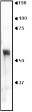
Fig.1 Western blot of tissue extract of mouse cerebral cortex with anti-nucleobinding 2 antibody (NET1).
Extract 10 μg protein was used. Antibody was used at 1/500 dilution.
The apparent molecular mass (55~57 kDa) is larger than the calculated one (50 kDa) and the band is broad, which may reflect post-translational modifications (one glycosylation and five phosphorylation sites)
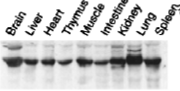
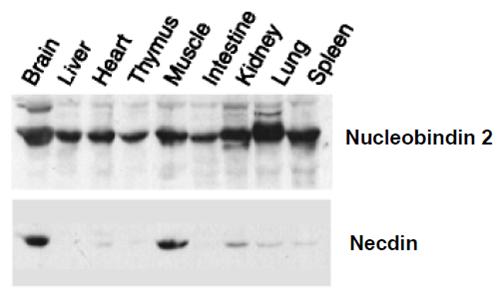
Fig.2 Expression of Nucleobindin-2 in various tissues as examined by western blotting.
Distribution of nucleobindin 2 in neonatal mouse organs. Homogenates of various organs from P0 mouse were separated by 10% SDS-PAGE and immunoblotted with this antibody. The antibody was used at 1/1,000 dilution.
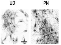
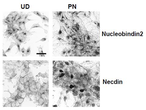
Fig.3. Immunocytochemistry for endogenous nucleobindin 2
Cells were stained with this antibody by the avidin-biotin-peroxidase complex method.
Left panel; undifferentiated murine embryonal carcinoma P19 cells (UD). Right panels, enriched post-mitotic neurons (PN). Nucleobindin 2 was localized to the cytoplasm near the nucleus in undifferentiated P19 cells, and its immunoreactivity in the cytoplasm was increased when P19 cells were induced to differentiate into neurons.
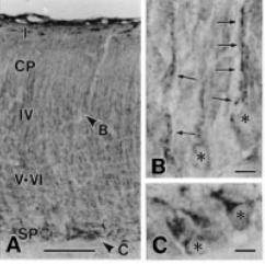
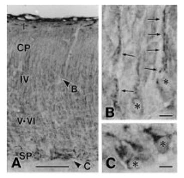
Fig.4 Immunohistochemistry for nucleobindin 2 in neonatal mouse brain with this antibody
Frozen brain sections from neonatal mouse were stained with this antibody by the avidin-biotin peroxidase complex method. A-C, cerebral cortex (parietal lobe). At higher magnification (B, C), fine granular immunoreactive materials are observed at both neuronal dendrites (arrows) and perikarya (asterisks) in the layer IV (arrowhead B in A) and subplate (arrowhead C in A) of the cerebral cortex.
Scale bars, 100 um (A) and 10um (B and C).
Reviews
There are no reviews yet.