

Description
p53 mutants are found in more than half of human cancers and are considered as the most important human cancer related gene. p53 is detected at 53kD position by electrophoresis and is composed of 393 amino acids. In the unstressed normal cells, the p53 level is low and it is inactive. However, with stress, especially with DNA damage, it is activated to promote arrest of cell cycle and repair of DNA damage, or induction of apoptosis. The functions and stability of p53 are regulated by phosphorylation of serine and threonine, and acetylation of lysine at various sites in the molecule. Ser315 is phosphorylated by aurora kinase and cycline-dependent kinases when cells are subjected to stress such as DNA damage and microtubule disruption by nocodazole (ref 1, 2 & 3). However the effect of the phosphorylation on the function of p53 is mostly unknown. This product is the purified IgG fraction obtained from serum-free culture medium of mouse hybridoma (clone #18) which produces monoclonal antibody that specifically recognizes human p53 protein with phosphorylated Ser315.
Applications
- Western blot x 1,000~2000 dilution, Fig.1
- Immunohistochemistry, Fig.2
- ELISA
Other applications have not been tested.
Specification
Antigen: synthetic peptide of Ser315-phosphorylated p53
Isotype: mouse IgG2bĸ
Form: purified monoclonal antibody (IgG) 1mg/ml in PBS (ph 7.4), 50% glycerol
Storage: -20 °C, for long term storage, -70°C
Data Link: UniProtKB/Swiss-Prot P04637 (P53_HUMAN)
References
- Katayama H et al “Phosphorylation by aurora kinase A induces Mdm2-mediated destabilization and inhibition of p53” Nature Genet. 36:55-62 (2004) PMID: 14702041
- Blaydes JP et al “Stoichiometric phosphorylation of human p53 at Ser315 stimulates p53-dependent transcription”J Biol Chem 276:4699-4708 (2001) PMID: 11078726
- Bode AM & Dong Z “Post-translational modification of p53 in tumorigenesis” Nature Rev Cancer 4: 793-805 (2004) PMID: 15510160
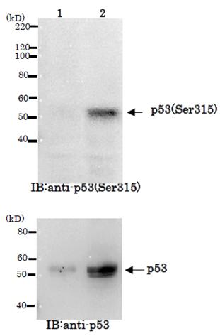
Fig.1 Identification of Ser315-phosphorylated p53 protein by Western blot. Sample: Crude cell extracts of MCF7 untreated (lane 1) and treated with nocodazole at 100 ng/ml for 48 h (lane2). The lower panel is the whole p53 protein identified by
omnipotent anti-p53 antibody (DO-1).
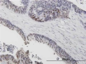
Fig. 2 Immunohistochemistry of stomach cancer. (Formalin/PFA-fixed paraffin-embedded section)
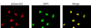
Fig.3. Immunofluorescence staining of p53 phosho-Ser315 in nuclei of HeLa cells subjected DNA damage. HeLa cells were treated with 100 nM Doxorubicin for 24 hr, fixed with 4% paraformaldehyde overnight, permealized with 0.25% Triton X-100 in PBS for 10 min. The cells were blocked prior to incubation with 1/1,000 diluted anti-p53(p-315) antibody in 1% BSA in PBS at 4℃ overnight. The cells were stained with secondary antibody, goat anti-mouse IgG conjugated with Alex 488, at 1/1,000 dilution in 1% BSA for 1 hr at room temperature. Nucleus (DNA) was stained with DAPI.
Reviews
There are no reviews yet.