

Description
Background:
PDILT cooperates with the testis-specific calreticulin-like chaperone, calsperin (CALR3), in the endoplasmic reticulum and plays an indispensable role in the disulfide-bond formation and folding of ADAM3. Pdilt(-/-) mice were male infertile because ADAM3 could not be folded properly and transported to the sperm surface without the PDILT/CALR3 complex.
Molecular mass: 67,759 with 588 amino acids. N-glucosylated
Specifications:
Reactivity: Mouse. Not tested in other species.
Validation: Specificity validated with knock-out mice.
Immunogen: Two synthetic peptides corresponding to the C-terminal regions of mouse PDILT, C+IRKPEEPERRKETA (550-563) and C+QPKEQPKPERKLEV (571-584), respectively, conjugated with KLH
Form: Whole rabbit antiserum added with 0.1% sodium azide.
Storage: Ship at 4℃and store at -20℃.
Applications:
- Western blotting (1/1,000 dilution)
- Immunoprecipitation (1/100)
- Immunohistochemistry (1/100-1/500)
Not tested for other applications.
Database Links: uniprot/Q9DAN1 Mouse PDILT, Gene ID 71830 mouse Pdilt
Reference: This antibody was described and used in the following publication.
- Tokuhiro K. et al (2012). Protein disulfide isomerase homolog PDILT is required for quality control of sperm membrane protein ADAM3 and male fertility. Proc Natl Acad Sci U S A. 109:3850-5. WB, IP. IHC. Free access.
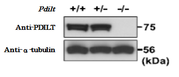
Fig. 1. Validation of specificity of the anti-PDILT antibody with knockout mice testis extracts. Samples wild-type, hybrid and knock-out were reacted with anti-PDILT antibody at 1/1,000 dilution. α-Tubulin was used as a control.
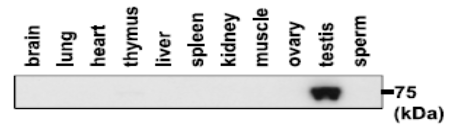
Fig.2 Western blotting analysis of PDILT expression in various tissues with anti-PDILT antibody.
Proteins were extracted from various tissues with lysis buffer containing Triton X-100 and subjected to western blot analysis. Tissue proteins (30 µg) and sperm protein (10 µg) were reacted with anti-PDILT antibody at 1/1,000 dilution.
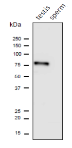
Fig.3 Western blot analysis of PDILT protein in the lysates of mouse testis and sperm with anti-PDILT antibody.
Proteins in the lysates (10 μg) were separated on SDS-PAGE, electro-blotted to PVDF membrane and reacted with anti-PDILT antibody at 1/1,1000 dilution. Anti-rabbit IgG conjugated with HRP was used at 10,000 dilution.
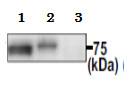
Fig.4. Immunoprecipitation of PDILT protein from mouse testis extracts. Extracts (100 μg) of wild-type (+/+) and Pdilt (-/-) mouse testes were immunoprecipitated with anti-PDILT antibody at 1/100 dilution and the precipitates were analyzed by western blotting using the same antibody at 1/1,000 dilution.
- Input. 2. Wild-type. 3. Pdilt (-/-)
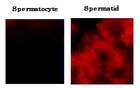
Fig.5. Immunofluorescence staining of mouse testicular sections with anti-PDILT antibody. Testes were collected from adult mice and were fixed in 4% paraformaldehyde/PBS, cryopreserved in garaded 10-30% sucrose, and embedded in a TissueTek OCT compound (Sakura Finetechnical, Tokyo). Frozen sections (5 μm) were mounted on APS-coated glass slides. After washing and blocking, the slides were reacted with anti-PDILT antibody at 1/100 dilution. As a secondary antibody, Alexa Fluor 546 conjugated anti-rabbit IgG antibody was used. PDILT was detected only in spermatid.
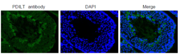
Fig.6 Immunohistochemical analysis of paraffin embedded mouse testis tissue labelling PDILT with PDILT antibody
Deparaffinization LemosolRA (#122-03991,Wako, Osaka)
Rehydration
Antigen retrieval Histo/Zyme (Cat.# k046; Diagnostic BioSystems)
Washing PBST (0.25% triton X-100/PBS-)
Blocking 10 % FBS / PBST 30 min
1st antibody 1/1,000 dilution in PBS- 4℃ O/N
Washing PBS-
2nd antibody 1,000 dilution, 60 min
washing PBS- 5 min X 3
DAPI 1.0μg/mL DAPI in TBS 10 min
Washing PBS-
Mount ImmunoSelect Antifading Mounting Medium (SCR-38447;
Reviews
There are no reviews yet.