

Description
Background:
PMIS2 might function both as the ultimate factor regulating sperm transport into the oviducts and in modulating sperm–zona binding.
Molecular mass: 11,040 with 96 amino acids. Likely to be N-glycosylated
Expression: Testis and sperm. Not expressed in other organs
Key words: Pmis2, Sperm, Fetilization,
Specifications:
Validation: Specificity has been validated with KO mouse (Fig.1)
Reactivity: mouse
Immunogen: Synthetic peptide corresponding to mouse PMIS2 (amino acids 61-80), CSNWEDAYRNSSRTMWFNML,conjugated with KLH
Form: Whole rabbit antiserum added with 0.1% sodium azide.
Storage: Shipped at 4℃ and store at -20℃.
Applications:
- Western blotting (1/1,000 dilution)
- Immunohistochemistry (1/1,000 dilution)
Database Links: uniprot/Q497Q9 mouse Pmis2.
Reference: This antibody was described and used in the following publication.
- Yamaguchi R. et al. (2012) Mice expressing aberrant sperm-specific protein PMIS2 produce normal-looking but fertilization-incompetent spermatozoa. Mol Biol Cell. 23:2671-9.
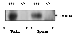
Fig. 1. Analysis of PMIS2 protein expression in the lysates of testis and sperm from wild-type (+/+) and Pmis2 knockout (-/-) mice by western blotting with anti-PMIS2 antibody. Testes and sperm were lyzed in lysis buffer containing 1% Triton-X100 and extracts were prepared as supernatants of lysates after centrifugation. Samples were reacted with anti-PMIS2 antibody at 1/1,000 dilution. The difference in predicted molecular mass (11 kDa) and the band position (18 kDa) of PMIS protein might be due to glycosylation.
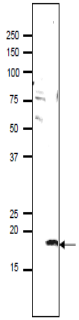
Fig.2 Identification of PMIS2 protein in the lysate of mouse sperm by western blotting with anti-PMIS2 antibody.
Proteins in the lysate was separated on SDS-PAGE (10~20% gradient gel), electro-blotted to PVDF membrane and incubated in anti-PMIS2 antibody at 1/1,000 dilution. As the second antibody, goat anti-rabbit IgG antibody conjugated with HRP (Abcam; ab97051) was used at 1/10,000 dilution.The numbers on the right are positions of molecular size markers in kDa.
The arrow indicates the position of PMIS2 protein.
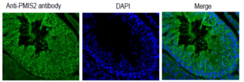
Fig.3 Immunohistochemical analysis of paraffin embedded mouse testis tissue with PDILT antibody
Deparaffinization LemosolRA (#122-03991,Wako, Osaka)
Rehydration by ethanol
Antigen retrieval Histo/Zyme (Cat.# k046; Diagnostic BioSystems, California))
Washing PBST (0.25% triton X-100/PBS-)
Blocking 10 % FBS / PBST 30 min
1st antibody 1/1,000 dilution in PBS- 4℃ O/N
Washing PBS-
2nd antibody Goat anti-rabbit IgG Alexa Fluor 488 conjugated,
1,000 dilution, 60 min
Washing PBS- 5 min X 3
DAPI 1.0μg/mL DAPI in TBS 10 min
Reviews
There are no reviews yet.