
Description
Description
The Rad6 (UBE2B)-Rad18 pair of genes plays a critical role in post-replication repair of damaged DNA. Rad6 protein functions as an E2 enzyme and Rad18 (509 aa, 57.4 kDa) as an ubiquitine ligase (E3) which ubiquitinates PCNA. Rad18 recruits translesion DNA polymerases to damaged DNA.
Applications (see Ref. 1-3)
- Western blot (1,000 fold dilution).
- Immunoprecipitation (200~500 dilution))
- Indirect immunofluorescence staining. (assay dependent)
- Immunohistochemistry (100~300 fold dilution)
Specification
Immunogen: GST-fusion protein containing 100 carboxyl terminal amino acids of mouse Rad18
Reactivity: Mouse Rad18 protein. Not reactive to human Rad18.
Product: IgG fraction of anti-mouse Rad18 rabbit serum
Form: 1 mg/ml in PBS, 50% glycerol, filter-sterilized, sodium azide and carrier-protein free
Storage: Ship at 4°C or -20°C. Upon arrival, centrifuge, and store at -20°C.
Data Link UniProtKB/Swiss-Prot Q9QXK2 Mouse Rad18
Gene ID 16098139 Mouse Rad18
References: This product has been used in the following publications.
- Tateishi S. et al. (2003) Enhanced genomic instability and defective post replication repair in RAD18 knockout mouse embryonic stem cells. Mol Cell Biol 23:474-81. PubMed 12509447 WB, IF/IC
- Watanabe K. et al. (2004) Rad18 guides poleta to replication stalling sites through physical interaction and PCNA monoubiquitination. EMBO J. 23:3886-96. PubMed 15359278 WB
- Masuyama S. et al. (2005) Regulated expression and dynamic changes in subnuclear localization of mammalian Rad18 under normal and genotoxic conditions. Genes Cells. 10:753-62. PubMed 16098139 IHC
- Sun J. et al. (2009) Rad18 is required for long-term maintenance of spermatogenesis in mouse testes. Mech Dev 126:173-83.PubMed 19068231 IHC, WB
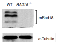
Fig.1.Identification of mouse Rad18 protein in ES cells by Western blot with anti-mRad18 antibody.
WT: Lysate of wild-type mouse ES cells
RAD18-/-: Lysate of Rad18 double knock-out mouse ES cells
Protein levels of α-tubulin in the lysates are shown as a control.
Three bands are absent in RAD18 knock-out cells.
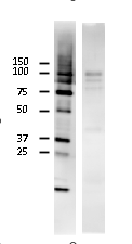
Fig.2. Identification of mouse Rad18 protein in NIH3T3 cells by Western blot with the antibody. Cell extract (23 ug) was used. Anti-mouse Rad18 was used at 2,000 fold dilution. Similar to Fig.1, two extra bands (75~90 kDa) may represent modified products (ubiquitination, phosphorylation).
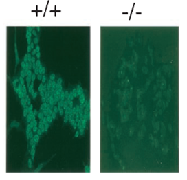
Fig.3. Immunofluorescence staining of Rad18 protein with anti-mRad18 antibody. Wild-type (+/+) and RAD18-/- ES cells (-/-). Samples were prefixed 3.7% formaldehyde and fixed with 80% methanol. Anti-mRad18 antibody was used at 1/300 dilution. As a second antibody, FITC conjugated anti-rabbit IgG was used.
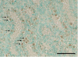
Fig.4. Detection of Rad18 in mouse testis.
Section of paraformaldehyde fixed mouse was stained with anti-mRad18 antibody. As a second antibody, peroxidase-conjugated anti-rabbit IgG donkey antibody was used. Signals were enhanced with TSA plus biotin system and detected by using DAB substrate.
Arrows indicate undifferentiated spermatogonia.
Reviews
There are no reviews yet.