
Description
Background:
Human Rad51 protein is a functional and structural homolog of E.coli RecA protein, which plays a major role in genetic recombination and recombination repair by mediating strand exchange reaction between homologous DNA strands (Ref.1). Rad51 functionally and physically interacts with its paralogs Dmc1, Rad51B, Rad51D, Xrcc2 and Xrcc3, and also with Rad52 in recombination processes. It also interacts with oncogene products and tumor suppressors such as BRACA1, BRACA2, and p53 for the maintenance of genome stability.
Specifications
Reactivity: human, rodents, chicken, frog (Xenopus)
Purity: Affinity-purified with full-size recombinant human Rad51 protein
Form:1 mg/ml in PBS with 50% glycerol, filter-sterilized. Azide-and carrier-free
Immunogen: Full-size recombinant human Rad51 protein (10-001)
Storage: Shipped at 4℃ and store at -20℃ (Do not store below -20℃ to avoid freezing)
Applications
- Western blotting (1/1,000 ~1/10,000 dilution). Fig. 1
- Immunoprecipitation (1/1,00-1/1,000). Fig. 2
- Chromatin Immunoprecipitation (ChIp) (assay dependent)
- Immuno-fluorescent staining (1/1,000~1/10,000). Fig. 3
- Immunohistochemistry (1/100-1/1,000). Fig.4
- Dot blotting (1/1,000~1/5,000 dilution)
- ELISA (indirect, 1/2,000~1/5,000 dilution)
Data Link UniProtKB/Swiss-Prot Q06609 (RAD51_HUMAN)
UniProtKB/Swiss-ProtQ08297 (RAD51_MOUSE)
References: Please let us know any publications using this antibody.
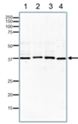
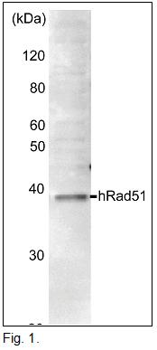
Fig.1 Western blotting of endogenous Rad51 proteins in crude extracts of varius animal cells.
15% SDS-PAGE was used. Blotting was done at 15 v overnight with wet system. The antibody was used at 1/1,000 dilution.
- MCF7 (human) cells. 40 μg
- NIH3T3 (mouse) cells. 40 μg
- CHO (hamster) cells. 40 μg
- Xenopus laevis (frog) egg. 40 μg
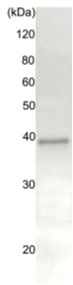
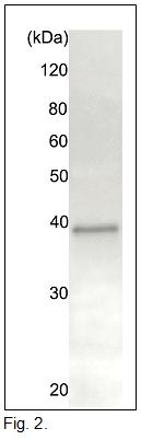
Fig.2. Immunoprecipitation of Rad51 protein from crude extract of HeLa cells by the anti-Rad51 antibody.
Anti-Rad51 antibody (20 ug) was incubated with 20 ug of HeLa cell extract, and precipitated with 20 ug of Pprotein A-beads. The sample was dissociated from the precipitate by heating in SDS-sample buffer and analyzed by western blotting with anti-Rad51 antiserum (chicken, ab63802) at 1/1,000 dilution. As second antibody, anti-chicken IgG antibody (rabbit) was used at 1/10,000 dilution.
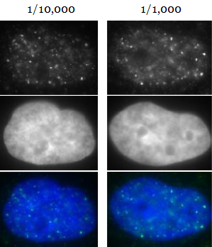
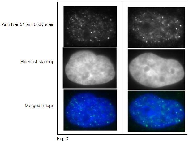
Fig.3. Detection of Rad51 foci formation after X-ray irradiation in human fibroblast cell line, GM0637.
Cells were irradiated by X-rays at 2 Gy,, grown for 1 hr, fixed with 4% paraformaldehyde in 1x PBS for 10 min, washed 3 times with PBS for 3 min, permealized by treatment with 0.5% Triton for 5 min, washed 3 times with PBS for 3 min, incubated with anti-Rad51 antibody for 30 min at 37℃, washed 3 times with PBS for 3 min, incubated with secondary antibody for 30 min at 37℃, washed 3 times with PBS for 3 min, stained with Hoechst for 1 min and mounted. Anti-Rad51 antibody was used at 1/10,000 dilution (left panels) and 1/1,000 dilution (right panels). As the secondary antibody, anti-rabbit Alexa 488 was used at 1/10,000 dilution. The pictures were by courtesy of Prof. S. Tashiro and Dr. K. Kono at Hiroshima University.
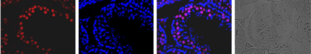
Fig.4. Immunohistological staining of Rad51 protein in mouse testis using anti-Rad51 antibody.
A section of formalin fixed and paraffin embedded mouse testis was treated with the anti-Rad51 antibody at 1/100 dilution after deparaffization and antigen retrieval. The 2nd antibody, anti-rabbit IgG conjugated with Alexa Fluor 647 (Abcam) was used at 1/1,000 dilution. DNA was stained with DAPI and the merged image was shown (Merged). The plain black and white microsopic picture of the same region was shown on the right.
Related Products:
Reviews
There are no reviews yet.