
Description
Background:
RanBPM (RanBP9) was identified as a protein that interacts with small GTP-binding protein, Ran, and forms a 670 kD multiprotein complex. The protein consists of 729 amino acids and is identified by Western blotting as an apparent molecular mass of 95 kD (see below). It is involved in nucleation of microtubules and controls cell growth by interacting with many protein factors.
Posttranslational modification: Ubiquitination. Phosphorylation.
Specificions:
Form: Undiluted antiserum added with 0.09% sodium azide
Validation: Indpendently validated in Ref.3 for IF.
Reactivity: human, monkey, rodents, dog
Immunogen: Recombinant human RanBP9 from Phe133 to Tyr 229.
Storage: Shipped at 4℃ and store at -20℃
Applications
- Western blotting (~1/ 2,000 dilution)
- Immunoprecipitation (assay dependent concentration)
- Immunofluorescence staining (1/200~1/1,000 dilution)
- Immunohistochemistry (1/200 dilution. Perform heat mediated antigen retrieval with citrate buffer (pH 6) before formalin treated paraffin embedded sectioning)
Data Link UniProtKB/Swiss-Prot Q96S59 (RANB9_HUMAN)
References
This product has been used in the following publications.
- Nishitani H et al “Full-sized RanBPM cDNA encodes a protein possessing a long stretch of proline and glutamine within the N-terminal region, comprising a large protein complex” Gene 272: 25-33 (2001) PMID: 11470507 WB, IP, IF (human, monkey, mouse, hamster)
- Umeda M et al ”A novel nuclear protein, Twa1, and Muskelin comprise a complex with RanBPM” Gene 303:47-54 (2003) PMID: 12559565 WB, IF (monkey)
- Madepalli K et al. “Role of RanBP9 on amyloidogenic processing of APP and synaptic protein levels in the mouse brain” FASEB J. 2012 May; 26(5): 2072–2083. PMID: 3336780 IF (mouse)
Salemi LM et al. “Aggresome formation is regulated by RanBPM through an interaction with HDAC6.” Biol Open. 2014 May 2;3(6):418-30. PMID: 24795145 WB, IP (human)
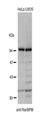
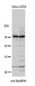
Fig.1 Identification of endogenous RanBP9 in crude extracts of HeLa and U2OS cells by Western blotting, using the anti-BP9 antibody. The antibody was used at 1/2,000 dilution.
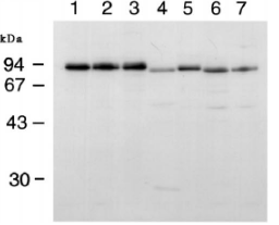
Fig.2 Western blotting of RanBP9 in various animal cells.
Whole cell extracts (50 ug) from human HeLa (1) and KB cells (2), Green Monkey Cos-7 cells (3), Chinese hamster CHO cells (4), mouse WEHI (6),and FM3A cells (7) were analyzed by western blotting with anti-BP9 antibody at 1/1,000 dilution.
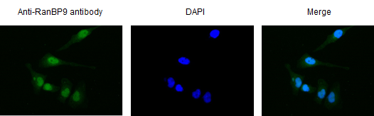
Fig.3 Immunofluorescece staining of RanBPM in HeLa cells by using anti-RanBPM antibody.
The cells were fixed with 4% PFA. The antibody was used at 1/1,000 dilution. As the secondary antibody, Alexa Fluor 488 conjugated goat anti-rabbit IgG antibody was used at 1/1,000 dilution. Nuclear DNA was stained with DAPI. RanBP9 is localized in perinuclear region.
Related product: Expression plasmid of GFP-RanBP9 cDNA is available upon request.
#71-001-1 pEGFP-C2-RanBPM
Reviews
There are no reviews yet.