
Description
RBP2 was originally identified as a retinoblastoma binding protein. It is also known as JARID1A (Jumonji, AT rich interactive domain 1A). RBP2 plays both negative and positive roles in RB-mediated transcriptional activation, depending on the kinds of genes and regulates differentiation by its function as an H3K4 histone demethylase (1, 2 & 3).
Applications
Western blot ~1 μg/ml
Immunofluorescence staining
Other applications were not tested.
Specification
Immunog: A synthetic peptide corresponding to human RBP2, amino acids 1416-1434.
Isotype: Mouse IgG2a kappa
Form: Purified monoclonal antibody (IgG) 1mg/ml in PBS, 50% glycerol, filter-sterilized
Specificity: Specific to human and mouse RBP2. Can detect endogenous levels of RBP2.
Storage: ship 4°C and store -20°C, for long term storage -70°C
Data Link UniProtKB/Swiss-Prot P29375 (KDM5A_HUMAN)
References
- Lopez-Bigas N et al “Genome-wide analysis of the H3K4 histone demethylase RBP2 reveals a transcriptional program controlling differentiation” Moll Cell 31: 520-530 (2008) PMID: 18722178
- Klose RJ et al “The retinoblastoma binding protein BRP2 is an H3K4 demethylase” Cell 128: 889-900 (2007) PMID: 17320163
- Christensen J et al “RBP2 belongs to a family of demethylases, specific for tri- and dimethylated lysine 4 on histone 3” Cell 128:1063-1076 (2007) PMID: 17320161
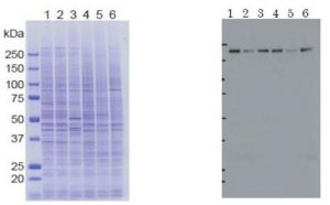
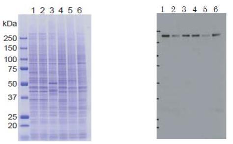
Fig. 1 Identification of RBP2 in crude cell extracts by Western blot with antibody 18E8. Samples: Lane 1: HeLa control siRNA, Lane 2: HeLa RBP2 siRNA, Lane 3: MCF7, Lane 4: U2OS, Lane 5: NIH3T3, Lane 6: J1 (mouse ES)
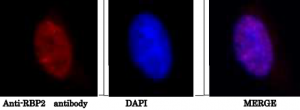
Fig. 2 Immunofluorescence staining of HeLa cell with anti-RBP” antibody.
- HeLa cells were fixed with 4% paraformaldehyde overnight, permealized with 0.25% Triton X-100 in PBS for 10 min. 2. Incubate cells with 1.5% BSA in PBS for 30 min to block non-specific binding of the antibodies. Incubate the cells with 1/2,000 diluted anti-RBP2 antibody (18E8) in 1% BSA in PBS at 4℃ overnight. 3. Incubate cells with a secondary antibody, goat anti-mouse IgG conjugated with Alex 488, at 1/1,000 dilution in 1% BSA for 1 hr at room temperature. 4. Nucleus (DNA) was stained with DAPI
Reviews
There are no reviews yet.