
Description
Background:
Ribonucleoside-diphosphate reductase subunit M2 (RRM2; 389 aa, 45 kDa) also known as ribonucleotide reductase subunit R2 (RNR-R2), is a rate-limiting subunit of an enzyme that catalyzes the formation of deoxyribonucleotides from ribonucleotides. Deoxyribonucleotides in turn are used in the synthesis of DNA. Furthermore, RNR plays a critical role in regulating the total rate of DNA synthesis so that DNA to cell mass is maintained at a constant ratio during cell division and DNA repair. It has been shown that MMR2 undergoes phosphorylation at Ser20 and Thr33.
Specifications:
Immunogen: Synthetic peptide (11 amino acids) in the C-terminal region of human and mouse RRM2, conjugated with KLH. The exact sequence is commercially sensitive.
Purity: Affinity-purified with the immunogen peptide
Form: 1mg/ml in PBS, 50% glycerol. Filter-sterilized. Azide and carrier free.
Reactivity: human, mouse, rat, hamster and Xenopus
Storage: Shipped at 4℃. Upon arrival, aliquot and store at -20℃
Applications
- Western blotting (1/1,000-1/2,000 dilution)
- Immunoprecipitation (1/300-1/1,000 dilution)
- Immunofluorescence staining (1/100-1/1,000 dilution)
- Immunohistochemistry; paraffin section (1/300 dilution)
Data Link UniProtKB/Swiss-Prot P31350 (RIR2_HUMAN)
Reference: This product was used in the following publication.
Takada S. et al. Identification of ribonucleotide reductase protein R1 as an activator of microtubule nucleation in Xenopus egg mitotic extracts. Mol Biol. Cell 11: 41734187 (2000) PMID: 11102516 WB
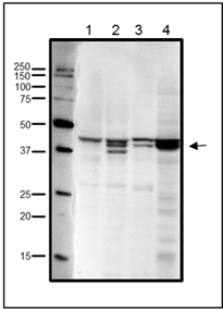
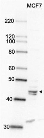
Fig.1 Western blot analysis of endogenous RRM2 in crude cell extracts. 1. HeLa cells (20 μg), 2. MCF7 cells (20 μg), 3. NIH3T3 cells (20 μg), 4. Xenopus eggs at mitotic stage (20 μg)
Multiple bands are due to phosphorylation at Ser20 and/or Thr33 (human sequence). The antibody was used at 1/1,000 dilution
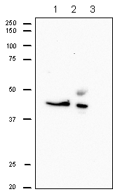
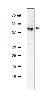
Fig.2 Immunoprecipitation of RRM2 from CHO cells.
Lane 1; Crude extract of CHO cells
Lane 2; The immunoprecipitate with the antibody at 1/1,000 dilution.
Lane 3; Supernatant of immuno –precipitation.
The upper band in lane 2 is IgG heavy chain. The antibody was used at 1/500 dilution
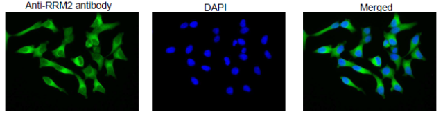
Fig.3 Immunofluorescence staining of RRM2 protein in MCF cells with anti-RRM2 antibody. MCF7 cells were fixed with 4%PFA and permeabilized with 0.25% TritonX 100 and reacted with anti-RRM2 antibody at 1/100 dilution. As the second antibody, anti-rabbit IgG antibody conjugated with Alexa Fluor 488 (Abcam) was used at 1/1,000 dilution. DNA was stained with 1.0μg/mL DAPI in TBS.
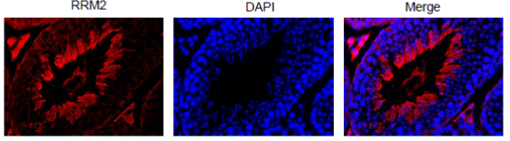
Fig.4 Immunohistochemical staining of RRM2 in mouse testis with anti-RRM2 antibody. Section of formalin-fixed and paraffin embedded mouse testis was reacted with anti-RRM2 antibody at 1/300 dilution. Nuclear DNA was stained with DAPI (center) and merged image is shown on left. RRM2 is abundantly expressed in actively proliferating cells.
Reviews
There are no reviews yet.