

Description
Background:
Component of AP2-containing clathrin coated structures at the plasma membrane or of endocytic coated vesicles. According to PubMed:15809293, probable serine/threonine-protein kinase that phosphorylates, in vitro, the beta2-subunit of the plasma membrane adapter complex AP2 and other proteins in presence of poly-L-lysine. According to PubMed:16914521, has no detectable kinase activity in vitro. May regulate clathrin-dependent trafficking between the TGN and/or the endosomal system.
Specifications:
Reactivity: Human, mouse, rat and hamster.
Validation: Specificity of reaction was validated with siRNA
Immunogen: Human SCYL2 protein (amino acids 528– 929) fused with a His6 tag
Purity: Purified IgG (Salting-out and ion-exchange chromatography)
Form: 1.0 mg/ml in 1 x PBS and 50% glycerol
Storage: Shipped at 4°C or -20℃ and store at -20℃
Applications
Western blotting (1/1,000 dilution)
Immunoprecipitation (1/200-1/1,000 dilution)
Immunofluorescence staining (1/200-1/1,000 dilution)
Data Link: UniProtKB Q6P3W7 (SCYL2_HUMAN)
Reference: This protein was described and used in the following publication.
- Terabayashi T. et al. A coated vesicle-associated kinase of 104 kDa (CVAK104) induces lysosomal degradation of frizzled 5 (Fzd5). J Biol Chem.(2009) 284(39):26716-24. WB, IP
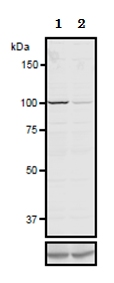
Fig.1 Validation of the anti-SCYL2 antibody with
siRNA.in western blotting.
293 cells were treated with control of SCYL2-siRNA. At 48 h after transfection,the lysates were analyzed by western blotting with anti-SCYL2 antibody or anti-β-tubulin antibody, the latter for a loading control.
Lane 1: Control siRNA
Lane 2: SCYL2-siRNA
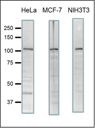
Fig.2. Detection of endogenous levels of SCYL2 in human and mouse cell extracts by western blotting.
20 μg of lysates of HeLa, MCF7 and NIH3T3 cells were used for western blotting. 7.5% gel was
used and blotted overnight in a wet system.
The anti-SCYL2 antibody was used at 1/1,000 dilution and as the 2nd antibody, goat anti-rabbit IgG (Abcam 97051) was used at 1/10,000 dilution.
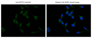
Fig.3 Immunofluorescence staining of SCYL2 in MCF7 cells.
MCF7 cells were fixed with 4% PFA and pemeabilized with 0.25% Triton X-100 in PBS.
The anti-SOYL2 antibody was used at 1/1,000 dilution and as a 2nd antibody, goat anti-rabbit IgG conjugated with Alexa Fluor 488 was used at 1/1,000 dilution (left panel). DNA was stained with DAPI (1 ug/ml) in TBS. The merged image was shown in the right panel.