

Description
Function: TAFIID subunit1 (1,872 aa, 213 kDa) is the largest component and core scaffold of the TFIID basal transcription factor complex. It contains novel N- and C-terminal Ser/Thr kinase domains which can autophosphorylate or transphosphorylate other transcription factors. It phosphorylates TP53 on ‘Thr-55’ which leads to MDM2-mediated degradation of TP53, and phosphorylates GTF2A1 and GTF2F1 on Ser residues. It possesses DNA-binding activity and is essential for progression of the G1 phase of the cell cycle.
Involvement in diseases: Dystonia 3, torsion, X-linked (DYT3) [MIM:314250]: A X-linked dystonia-parkinsonism disorder. Dystonia is defined by the presence of sustained involuntary muscle contractions, often leading to abnormal postures. DYT3 is characterized by severe progressive torsion dystonia followed by parkinsonism. It has a well-defined pathology of extensive neuronal loss and mosaic gliosis in the striatum (caudate nucleus and putamen) which appears to resemble that in Huntington disease.
Applications
- Western blotting (1/1,000 dilution)
- Immunofluorescent staining (1/1,000~1/5,000)
Other applications have not been tested
Specification
Immunogen: Synthetic peptide of human TFIID subunit 1 protein corresponding to amino acids 103-123, STEDAVDYSDINEVAEDESRR-C, conjugated with KLH
Reactivity: Human, hamster, mouse and rat. Not tested in other species.
Product: Rabbit whole antiserum added with 0.05% sodium azide
Storage: Shipped at 4°C. Upon arrival, spin-down and store at -20°C.
Data Link: uniprot/P21675 (TAF1_HUMAN)
Reference: This antibody has been used in the following publication:
Sekiguchi T. et al. (1991) The human CCG1 gene, essential for progression of the G1 phase, encodes a 210-kilodalton nuclear DNA-binding protein. Mol Cell Biol. 11: 3317-25 pubmed/2038334
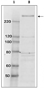
Fig.1 Identification of TAF1 protein in whole cell extract of HeLa cells by western blotting using anti-TAF1 antibody
Lane 1; Size marker proteins (kDa)
Lane 2; HeLa cell whole extract (10 μg)
Arrow indicates the position of TAF1 protein band
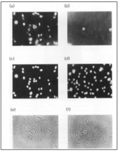
Fig.2. Immunofluorescence staining of TAF1 protein in BHK cells with anti-TAF1 antibody
(a) Reacted with anti-TAF1 antibody
(b) Reacted with anti-TAF1 antibody in the presence of the immunogen peptide.
(c) The same field as (a), stained with Hoechst 33258.
(d) The same field as (b), stained with Hoechst 33258.
(e) The same field as (a) and (c), photographed through phase-contrast microscope
(f) The same field as (b) and (d), photographed through phase-contrast microscope.
Growing cells were fixed with 3% formaldehyde and permeabilized 1% Nonidet P-40. As the 2nd antibody, goat anti-rabbit IgG conjugated with rhodamine was used. TAF1 protein was detected in nucleus.
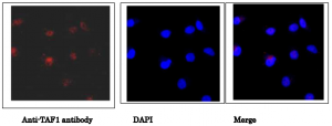
Fig.3. Immunofluorescence staining of TAF1 protein in HeLa cells with anti-TAF1 antibody
Hela cells were fixed with 4% paraformaldehyde and permeabilized with 0.25% Triton X-100. Anti-TAF1 antibody was used at 1/5,000 dilution. As a 2nd antibody, goat anti-rabbit IgG conjugated with Alex 488 was used at 1/1,000 dilution. Nucliei were stained with DAPI. The merged image was shown in the right panel. TAF1 protein was detected in nucleus.
Reviews
There are no reviews yet.