

Description
Background:
In Brassicaceae, the enzyme myrosinase (beta-thioglucoside glucohydrolase, TGG) degrades glucosinolates to produce toxins like thiocyanates, isothiocyanates, nitriles, epithionitriles or oxazolidine-2-thiones that deter herbivory. There are two TGG enzymes, TGG1 and TGG2, which have a redundant function.
Subcellular location: Vacuole
Modification: N-linked glycosylation at 9 asparagine residues. Elimination of 19-amino acid signal peptide from N-terminus.
Specifications:
Storage: Shipped at 4°C and store at -20℃
Form: 2 mg/ml in PBS, 50% glycerol. Filter sterilized. No preservative or carrier added.
Purity: IgG, affinity-purified with Protein A
Immunogen: A synthetic peptide, AQNNQTIVPSDVHT, corresponding to TGG1 protein (353-366) of A. thaliana, conjugated with bovine serum albumin.
Reactivity: TGG1 protein of Arabidopsis thaliana. Not tested for other species.
Validation: Specific reactivity has been validated by western blot showing that the TGG2 specific band is absent in tgg2-1 mutant leaf extract (Ref.1)
Applications
- Western blot (1/1,000- 1/3,000 dilution)
- ELISA (assay dependent)
- Immunohistochemistry (1/500-1/1,000)
- Immunoelectron microscopic analysis (1/1,000-1/2,500)

Fig. 1 Western Blot of TGG1 in Arabidopsis leaf extract.
Anti-TGG1 antibody was used at 1/1,000 dilution. Secondary antibody (goat anti-rabbit IgG antibody HRP-conjugated, ab97051) was used at 1/10,000 dilution.
Sample: Arabidopsis leaf extract, 10 μg
Molecular mass of TGG1 is 61 kDa from the amino acid sequence. The protein undergoes modifications such as elimination of signal peptide and glycosylation at 9 positions, which changes molecular mass in mature form.
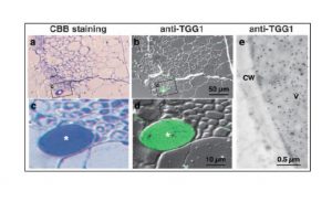
Fig. 2 Localization of TGG1 in sections of Arabidopsis rosette leaves
Sections of rosette leaves of 48-day—old plants were stained with CBB (a, c), and reacted with anti-TGG1 antibody at 1/1,000 dilution followed by reaction with Alexa Fluor 488 goat anti-rabbit IgG at 1/1,000 dilution (b, d). Images c and d are enlarged images of the boxed area in images a and b, respectively. Asterisks show myrosin cells.
For immunoelectron microscopy (e), ultrathin sections were mounted on Formvar-coated nickel grid. The sections were reacted with anti-TGG1 antibody at 1/1,000 dilution. After washing with PBS, they were incubated with anti-rabbit IgG conjugated to gold particle (AuroProbe EM). CW is cell wall and V, vacuole.