


Description
The VRK1 gene encodes serine/threonine kinase VRK1 (Vaccinia-Related Kinase 1; 396 aa, 45.5 kDa) which is involved in Golgi disassembly during the cell cycle following phosphorylation by PLK3 during mitosis, and required to induce Golgi fragmentation. It acts by mediating phosphorylation of a downstream target protein ‘Thr-18’ of p53/TP53 and may thereby prevent the interaction between p53/TP53 and MDM2. It also phosphorylates casein and histone H3. Phosphorylation of the BANF1 gene product disrupts its ability to bind DNA, reduces its binding to LEM domain-containing proteins and causes its relocalization from the nucleus to the cytoplasm.
Defects in VRK1 are the cause of pontocerebellar hypoplasia type 1A (PCH1A); also called pontocerebellar hypoplasia with infantile spinal muscular atrophy or pontocerebellar hypoplasia with anterior horn cell disease. PCH1A is characterized by an abnormally small cerebellum and brainstem, central and peripheral motor dysfunction from birth, gliosis and anterior horn cell degeneration resembling infantile spinal muscular atrophy.
Applications
- Western blot (1/200~1/1,000 dilution). Highly sensitive chemiluminescence reagents such as Lumi-Light Plus (Roche), ImmunoStarRLD, or ImmunoStarR Zeta (Wako, Osaka) are recommended.
- Immunoprecipitation (assay dependent)
- Immunofluorescence staining (1/100 dilution)
- Immunohistochemistry (assay dependent)
- ELISA (assay dependent)
Specification
Reactivity: Human VRK1 protein. Not tested with other species.
Immunogen: Synthetic peptide corresponding to N-terminus of human VRK1, MPRVKAAQAGRQSSAKRHL-C
Product: Mouse monoclonal antibody (5D1) produced in serum-free medium and purified by propriety chromatography under mild conditions (90~98% pure).
Isotype: IgG1, kappa
Form: 1 mg/ml in PBS with 50% glycerol, filter-sterilized.
Storage: Shipped at 4°C and store at -20°C
Data Link: SwissProt: Q99986 Human
Entrez Gene: 7443 Human
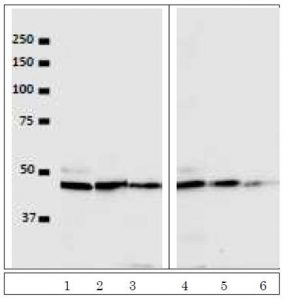
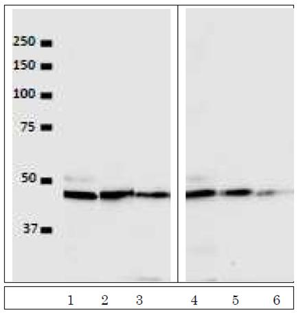
Fig. 1. Western Blot detection of VRK1 in the crude extracts of human cells. Lanes 1, 2, 3: HeLa cell extract (5×10⁴ cells) with antibody dilutions at 1/100, 1/500, 1/1000. Lanes 4, 5, 6: U2OS cell extract (5×10⁴ cells) with the antibody dilutions at 1/100, 1/500, 1/1000. As secondary antibody, Alexa488 conjugated
goat anti-mouse IgG was used. ImmunoStarR
LD (Wako, Osaka) was used as
Chemiluminescence reagent and images were taken with BIO-RAD ChemiDocXRS.
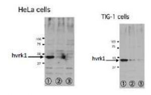
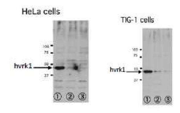
Fig. 2. Inhibition of VRK1 Expression in human cells by RNAi specific to VRK1.
Lane 1: Luciferase RNAi (control). Lane2: VRK1-1 RNAi. Lane 3:VRK1-2 RNAi Antibody at 1/500 dilution. Lumi-Light Plus (Roche) was used as chemiluminescence reagent. Extracts from 5×10⁴ cells.
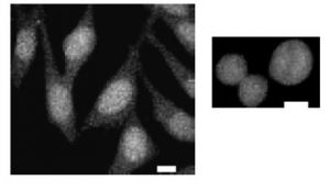
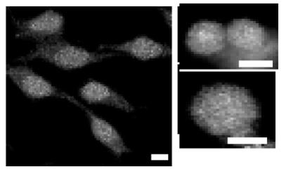
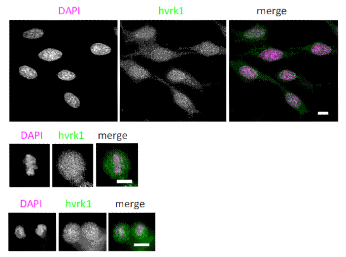
Fig. 3. Immunoflorescence staining of
VRK1 in HeLa cells. Left: Interphase cells fixed with paraformaldehyde and stained with the antibody at 1/100 dilution. Right: Metaphase cells. At metaphase, VRK1 dots were detected solely in nuclei.

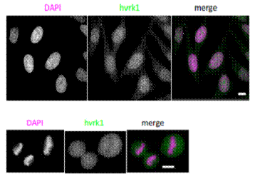
Fig. 4. Immunofluorescence staining of
VRK1 in Hela cells. Left: Interphase cells fixed with methanol and stained with the antibody at 1/100 dilution. Right: Metaphase cells.
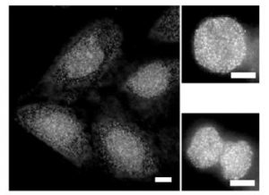
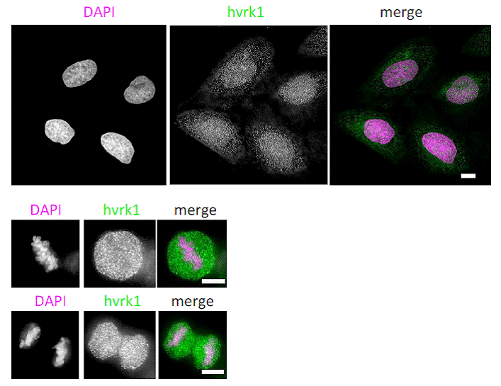
Fig. 5. Immunofluorescence staining of
VRK1 in U2OS cells. Left: Interphase cells fixed with paraformaldehyde and stained with the antibody at 1/100 dilution. Right: Metaphase cells.
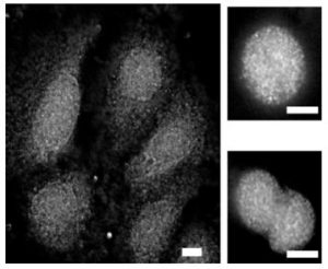
Fig. 6. Immunofluorescence staining of VRK1 in U2OS cells. Left:
Interphase cells fixed with methanol and stained with the antibody at 1/100 dilution. Right: Metaphase cells
Figures are provided by courtesy of Prof. Tokuko Haraguchi and Ms. Takako Kojin at Advanced ICT Institute, Kobe, Japan
Reviews
There are no reviews yet.