
Description
XP (Xeroderma pigmentosum) is an autosomal recessive human disease characterized by hypersensitivity to sunlight and a high incidence of skin cancer on sun-exposed skin (1). Cells from XP patients are hypersensitive to killing by UV irradiation because of a defect in nucleotide excision repair (NER). XP is classified into seven complementation groups (A~G) and a variant form (1). XPA shows the most severe symptoms. Products encoded by the XP genes function in repairing UV-induced cyclobutane pyrimidine dimmer and (6-4) photoproducts as well as chemically induced variety of DNA lesions (1).
XPA protein consists of 273 amino acids and forms a complex with many proteins such as RPA, ERCC1, TFIIH、XAB1, and XAB2, which plays a role in early step of NER. The hybridoma 5F12 was constructed by Prof. K. Tanaka’s group who first cloned the XPA gene (2, 3).
Applications
- Western blot 1:1000-10000 dilution
- Immunfluorescence staining 1:100-1000
- ELISA
- Inhibition of in vitro excision repair reaction
- Inhibition of XPA interaction with ERCC1 and TFIIH
Other applications have not been tested.
Specifications
Immunogen: Recombinant full-length human XPA protein
Reactivity: human (expected to react also with mouse XPA from the
sequence homology)
Epitope: Amino acids 30-47
Clone: Mouse monoclonal antibody, 5F12
Subtype: IgG2b
Form: Purified IgG, 1 mg/ml in PBS pH 7.2, 50% glycerol, filter-sterilized
Storage: Ship 4°C, upon arrival aliquot and store -20°C
Data Link
UniProtKB/Swiss-Prot P23025 (XPA_HUMAN)
References: This antibody is described in Ref. 2
- Friedberg EC et al DNA Repair and Mutagenesis 2nd ed., ASM Press (2006)
- Saijo M et al “Inhibition of nucleotide excision repair by anti-XPA monoclonal antibodies which interefere with binding to RPA, ERCC1, and TFIIH” Biochem Biophys Res Comm 321:815-822 (2004) PMID: 15358100
- Tanaka K et al “Analysis of a human DNA excision repair gene involved in group A xeroderma pigmentosum and containing a zinc-finger domain” Nature 348:73 -76 (1990) PMID: 2234061
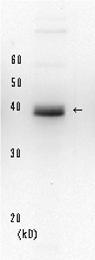
Figure 1 Detection of XPA protein in the crude extract of HeLa cells by Western blotting using this monoclonal antibody.
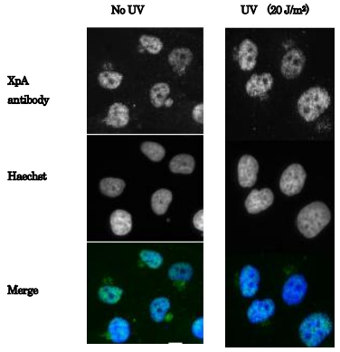
Fig. 2 Immunofluorescence staining of human fibroblast cells (GM0637) using anti-XpA antibody (5F12). The cells were non-irradiated (left) or irradiated with UV at 20 J/m2 and fixed after 30 min with paraform aldehyde. The antibody was used at 1/100 dilution and as the second antibody, Alexa 488 conjugated goat anti-mouse IgG was used at 1/5,000 dilution.
Reviews
There are no reviews yet.