
Description
Calmegin plays an important role in sperm fertility. Binds calcium ions. Specifically expressed during male meiotic germ cell development.
Molecular mass: 69,431 with 611 amino acids
Key words: Calmegin, CLGN, Spermatogenesis, Endoplasmic reticulum, Chaperon, Transmembrae, PDILT, Calcium ion binding, Binding of sperm to zona pellucida
Specifications:
Validation: Specificity validated with knock-out mouse (Fig.2.)
Reactivity: Mouse.
Immunogen: C-terminal peptide of mouse Calmegin, DESPGSGDAPLKSLRKRRVRKD, conjugated with KLH
Form: Whole rabbit antiserum added with 0.1% sodium azide.
Storage: Shipped at 4℃ and store at -20℃.
Applications:
Western blotting (1/1,000 dilution))
Immunoprecipitation (1/100~1/1,000 dilution).
Immunofluorescent staining (1/100~1/1,000 dilution)
Immunohistochemistry (Paraffin embedded) (1/1,000 dilution)
Database Links:
uniprot/P52194 mouse Calmegin
Gene ID 12745 mouse Clgn
Reference: This antibody was described in Ref.1 and used in the following publications.
Ikawa M. et al. (2001) Calmegin Is Required for Fertilin α/β Heterodimerization and Sperm Fertility. Dev Biol.240: 254-61. WB, Open access.
Ikawa M. et al. (2011) Calsperin is a testis-specific chaperone required for sperm fertility. J Biol Chem. 286: 5639-46. WB, IP. Open access.
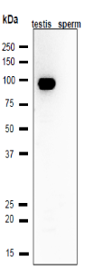
Fig.1 Western blot analysis of Calmegin in lysates of mouse testis and sperm. Proteins in the lysates (10 μg) were separated on SDS-PAGE (10~20% gel) and blotted to PVDF membrane. It was reacted with anti-Calmegin-antibody at 1/1,000 dilution. As the second antibody, goat anti-rabbit IgG conjugated with HRP (Abcam; ab97051) was used at 1/10,000 dilution.
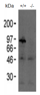
Fig.2 Western blotting analysis of testis extracts of wild-type and knockout mice with anti-Calmegin antibody. 20 μg of Triton X-100 extracts from mouse testes was reacted with anti-Calmedin antiserum at 1/1,000 dilution. Arrow indicates the position of intact Calmegin.
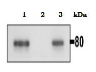
Fig.3. Immunoprecipitation of Calmegin from mouse testis. One mg of testis lysate was incubated with 2 μl of anti-Calmegin antiserum and 50 μl. of protein-A conjugated magnetic beads (Miltenyi Biotec) and immunoprecipitated according to the protocol of supplier. The immunoprecipitated sample was analyzed by western blotting with the antibody at 1/1,000 dilution.
1.Input 2. non-immune serum 3. Anti-Calmegin antiserum
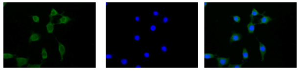
Fig.4 Immunofluorescent staining of Calmegin in NIH3T3 cells with anti-Calmegin antibody.
Fixation of the cells in 4% paraformaldehyde overnight
Permeabilization in 0.25% Triton X-100/PBS for 10 min
Blocking in 1.5% BSA/PBS for 30 min
1st antibodies 1/100 diluted by blocking buffer over night
2nd Goat anti rabbit IgG conjugated with Alex 488 (1:1000 dilution) for 60min
Nuclei were stained with DAPI
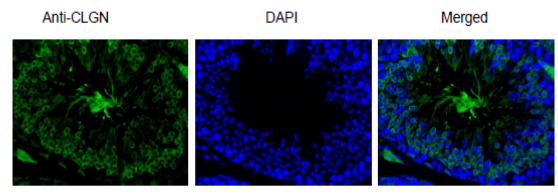
Fig.5 Immunohistological staining of Calmegin in testis section with anti-Calmegin antibody
Deparaffinization LemosolRA (#122-03991, Wako, Osaka)
Rehydration
Antigen retrieval Histo/Zyme (Cat.# k046; Diagnostic BioSystems)
Washing PBST (0.25% triton X-100/PBS-)
Blocking 10 % FBS / PBST 30 min
1st antibody 1/1,000 dilution in PBS- 4℃ O/N
Washing PBS-
2nd antibody Goat anti rabbit IgG conjugated with Alex 488 (1:1000 dilution) for 60min
Washing PBS- 5 min, 3 times
DAPI 1.0μg/mL DAPI in TBS 10 min (×100 stock, 0.1mg/mL)
Washing PBS-
Mount ImmunoSelect Antifading Mounting Medium (SCR-38447; Dianova
Reviews
There are no reviews yet.