
Description
E2F1 is a member of E2F group of proteins that share common structural and functional domains and plays a major role during the G1/S transition in the mammalian cell cycle as a transcriptional factor (1). E2F1 is regulated during cell cycle progression. It is phosphorylated at Ser364 by Chk2 kinase in response to DNA damage, stabilized, mobilized to nucleus and activated as a transcription factor (2). It induces apoptosis by activating transcription of the p53 homolog, p73 (3). E2F1 protein consists of 437 amino acids with a molecular mass of 46 kDa.
Applications
- Western blot ~1 µg/ml
- ELISA
Not tested for other applications.
Specification
Product: Mouse monoclonal antibody (clone #2) specific to the human E2F1 protein phosphorylated at Ser364. Produced in serum-free medium and purified under mild conditions
Antigen: A synthetic peptide corresponding to a sequence of human E2F1 protein including and surrounding phospho-Ser364
Isotype: IgG2b kappa
Form: Purified IgG 1 mg/ml in PBS(-), 50% glycerol
Reactivity: Human E2F1 protein phosphorylated at Ser364. Not tested with other species.
Storage: -20°C, for long term storage -70°C
Data Link: UniProtKB/Swiss-Prot Q01094 (E2F1_HUMAN)
References
- Trimarchi JM & Lees JA ”Sibling rivalry in the E2F family” Nat Rev Mol Cell Biol 3:11-20(2002) PMID: 11823794
- Stevens C et al “Chk2 activates E2F-1 in response to DNA damage” Nat Cell Bio1 5:401-409 (2003) PMID: 12717439
- Irwin M et al “Role for the p53 homologue p73 in E2F-1-induced apoptosis” Nature 407:645-648 (2000) PMID: 11034215
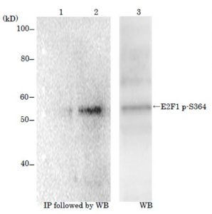
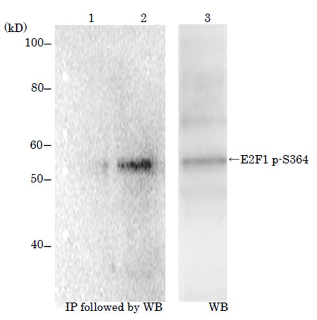
Figure: Identification of E2F1 protein phosphorylated at p-Ser364 with monoclonal antibody (#2)
MCF cells were grown in the absence (lane 1) or in the presence of etoposide at 10 μM for 16 h (lanes 2 & 3). Crude lysates were prepared and analyzed by Western blot (lane 3) with the anti-body #2 or immunoprecipitated by pantropic anti-E2F1 antibody followed by Western blot with the antibody #2.
Reviews
There are no reviews yet.