
Description
Hepatitis C virus (HCV) is a small (55-65 nm in size), enveloped, positive sense single-stranded RNA virus in the family Flaviviridae and the principal cause of parenteral non-A, non-B hepatitis. The virus genome consists of a single open reading frame of approximately 9.4 kb which encodes a single polyprotein of about 3,010 amino acids (1, 2, 3). The polyprotein is processed by host cell and viral proteases into four structural proteins (core, envelope 1 and 2, and p7) and six non-structural proteins (NS2, 3, 4a, 4b, 5a, and 5b) necessary for viral replication. HCV core protein (191 aa) is not only a component of nucleocapsid but also has multiple functions and is thought to be a pathogenic factor for hepatitis. It also participates in some cellular processes, including transcriptional regulation and cellular transduction. HCV core antigen is used as diagnostic marker for HCV infection.
Applications
- Western blot 2. Immunohistochemistry 3. Immunofluorescence staining
- ELISA 5. FACS
Specification
Immunogen: A part of the core region (nucleotides 369-704, amino acids 13-124) of HCV genotype 1b expressed in E. coli (the nucleotide sequence is shown in ref.3)
Conjugate: FITC conjugated, [FITC] / [IgG] = 6.7
Isotype: Mouse IgG2a kappa
Form: 1.6 mg/ml in PBS, 50% glycerol, filter-sterilized
Specificity: Specific to human HCV core antigen of genotype 1b. Not tested in other genotypes
Storage: Ship at 4°C and long term storage at -20°C
Data Link Swiss-Prot HCV protein
References: This antibody (unconjugated) has been used in ref.4 and 5.
- Brass V, Moradpour D, Blum HE. Molecular Virology of Hepatitis C Virus (HCV): 2006 Update. Int J Med Sci 2006; 3:29-34. PMID: 16614739
- Kato, N. et al. (1990) “Molecular cloning of the human hepatitis C virus genome from Japanese patients with non-A, non-B hepatitis.” Proc. Natl. Acad. Sci. USA 87, 9524-9528 PMID: 2175903
- Takamizawa, A. et al. (1991) “Structure and organization of the hepatitis C virus genome isolated from human carriers.” J.Virol.65, 1105-1113 PMID: 1847440
- Manabe, S. et al. (1994) “Production of nonstructural proteins of hepatitis C virus requires a putative viral protease encoded by N3.” Virology 198, 636-644 PMID: 8291245
- Hiramatsu, N. et al. (1992) “Immunohistochemical detection of hepatitis C virus-infected hepatocytes in chronic liver disease with monoclonal antibodies to core, envelope and NS3 regions of the hepatitis C virus genome.” Hepatology, 16, 306-311 PMID: 1379209
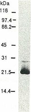
Fig. 1 Western blot of HCV core protein.
Chimp liver cells were infected with recombinant vaccinia virus containing a HCV genome cDNA and were subjected to Western blot using this antibody. The core protein is detected as a 22-kDa band.
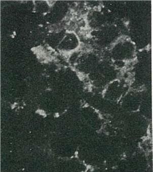
Fig. 2 Detection of HCV core protein by immuno-fluorescence antibody staining. Chimp liver cells were infected with recombinant vaccinia virus containing a HCV genome cDNA. After incubation for 48 hr, the cells were fixed with acetone and HCV core protein was detected by indirect immunofluorescence staining using this antibody.
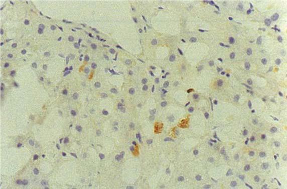
Fig. 3 Immunohistochemical detection of HCV core protein.
Tissue section from a patient with chronic hepatitis C was immunostained to reveal cells expressing HCV core antigen, which are scattered in the lobules (indirect immuno-histochemical method, counterstained with Mayer’s hematoxylin).
Reviews
There are no reviews yet.