
Description
Background:
DSN1 / Mis13 (human: 356 aa. 40 kDa) is part of the MIS12 complex which is required for normal chromosome alignment and segregation and kinetochore formation during mitosis. Defects in kinetochore proteins often lead to aneuploidy and cancer. It is a component of the MIS12 complex composed of MIS12, DSN1, NSL1 and PMF1 and also interacts with CASC5, CBX3 and CBX5. Note that DSN1 protein is expressed in actively dividing cells.
Specifications:
Product: Rabbit antiserum added with 0.05% sodium azide. Anti-GST antibodies have been removed from the serum with affinity column conjugated with GST
Validation of Reactivity: Specificity has been validated with siRNA of human DSN1 for WB and IF. Reacts with mouse, rat
Immunogen: Recombinant GST-DSN1 (1-356 aa)
Storage: Shipped at 4℃ and store at -20℃.
Applications:
- Western blotting (1,000-fold dilution)
- Immunofluorescence-staining (1,000-fold dilution,)
Data Link: UniProtKB/Swiss-Prot Q9H410 (DSN1_HUMAN)
Reference: This antibody has been used in the following reference
- Obuse C et al “A conserved Mis 12 centromere complex is linked to heterochromatic HP1 and outer kinetochore protein Zwint-1“ Nat Cell Biol 6: 1135-1141 (2004) PMID: 15502821
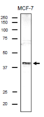
Fig.1 Identification of endogenous DSN1 protein in the crude lysate of MCF-7 cells by using anti-DSN antibody. The cell lysate (20 μg) was separated by SDS-PAGE and analyzed by western blotting with anti-DSN antibody at 1/1,000 dilution. As the second antibody, anti-rabbit IgG antibody conjugated with HRP (Abcam, ab97051) was used at 1:10,000 dilution.
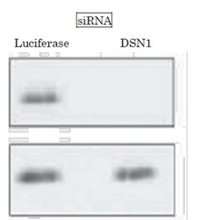
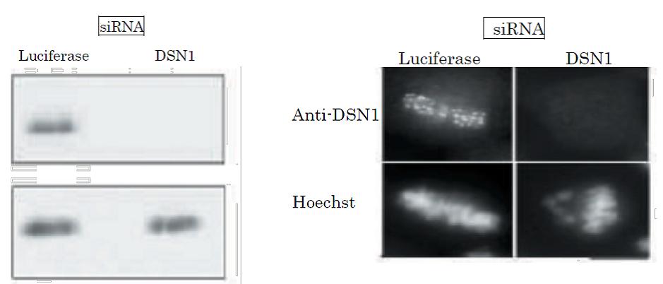
Fig.2 Validation of specificity of the anti-DSN1 antibody in WB by using siRNA. Expression of siRNA for DSN1 for 24 h greatly reduced DSN1 in the cells. Luciferase siRNA was used as a control. Antibody was used at 1/1,000 dilution.
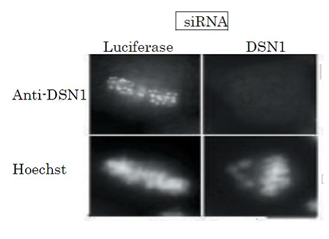
Fig.3 Immunofluorescence staining of HeLa cell chromosome with this antibody and validation of specificity with siRNA. Expression of siRNA for DSN1 for 24 h greatly reduced DSN expression in the cell. Luciferase siRNA was used as a control. Antibody was used at 1/1,000 dilution. Mitotic cells were accumulated by nocodazole treatment and chromosomes were spread on cover glass.
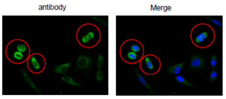
Fig.4 Immunofluorescence staining of DSN1 in HeLa cells using anti-DSN1 antibody. The cells were fixed with 4% paraformaldehyde. The antibody was used at 1/100 dilution. As the second antibody, Alexa Fluor conjugated goat anti rabbit IgG was used at 1/1,000 dilution. Nuclear DNA was stained with DAPI and the merged image was shown on the right.
Note that DSN1 is abundantly expressed in mitotic cells on mitotic apparatus.
Reviews
There are no reviews yet.